

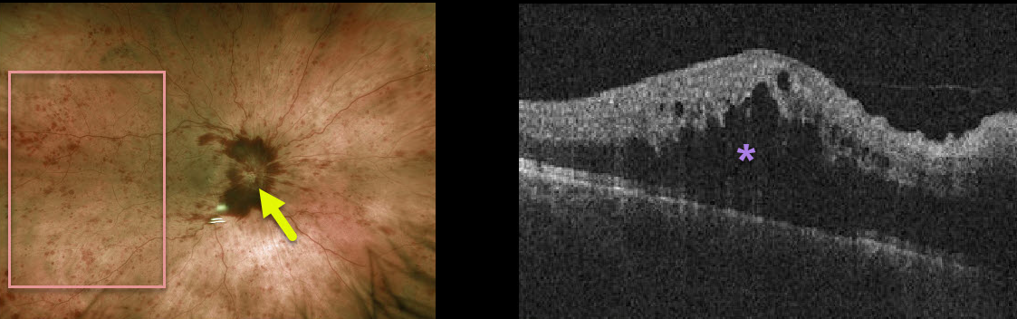
This patient has a non-ischemic central retinal vein occlusion (CRVO) with macular edema.
The fundus photo shows diffuse intraretinal hemorrhages, disc hemorrhages, and optic disc edema of the right eye. The OCT demonstrates marked macular edema.
Learning Points:
About 10% of patients with CRVO will develop bilateral occlusions. Eighty percent of occlusions are non-ischemic, although about one-third of these will convert to ischemic occlusions within several years.
About 60% of ischemic occlusions will develop anterior segment neovascularization with neovascular glaucoma. For some reason, posterior segment neovascularization rarely occurs (see the classic Hayreh 1983 paper Ophthalmology 1983;90;488-506).



