

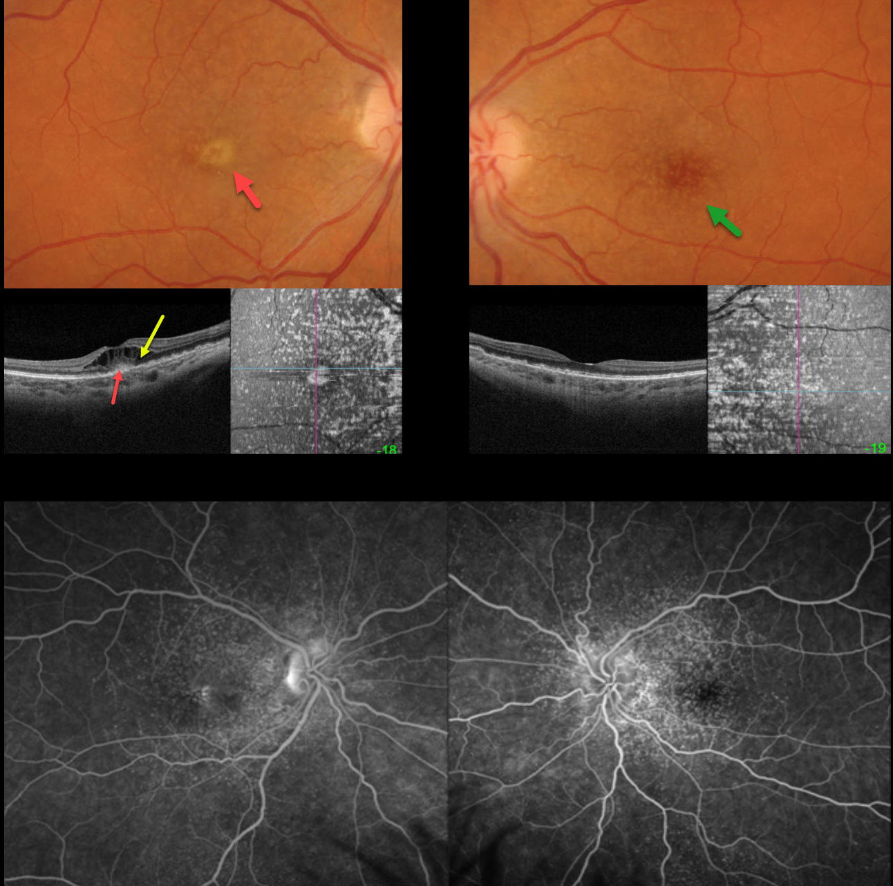
This 47YO female presented with one week of vision loss in her right eye. Vision was counting fingers OD and 20/25 OS.
Optos color RGB imaging shows innumerable cuticular macular drusen, which appear on swept-source OCT B-scanning as arrowhead-like RPE elevations and on en face imaging as numerous hyperreflective spots. A yellow subfoveal lesion OD appears on OCT as hyperreflectivity in the outer retina. Marked overlying cystoid edema is seen. Fluorescein angiography shows late pooling in the foveal cysts OD, with hyperfluorescent window defects from the drusen.
One month following an initial intravitreal Avastin injection, vision improved to 20/80, with complete resolution of the vitelliform lesion and cystoid edema. Treat-and-extend treatment was started.
Learning Points:
Originally described by Gass as basal laminar drusen (AJO 1985;99:445-459), cuticular drusen were initially thought to represent nodular thickening of the RPE basement membrane. They appear clinically as numerous, round, yellow-white ‘hard drusen’, and on OCT as small, arrowhead-shaped drusen located above Bruch’s membrane and below the RPE (Spaide and Curcio, Retina 2010;30:1441-1454).
Our patient’s angiographic appearance echoes Dr. Gass’ original description of “a beautiful stars-in-the-sky or Milky-Way pattern” of hyperfluorescence. Cuticular drusen are classically associated with avascular vitelliform lesions (Finger et al, Retina 2010;30:1455-1464).



