

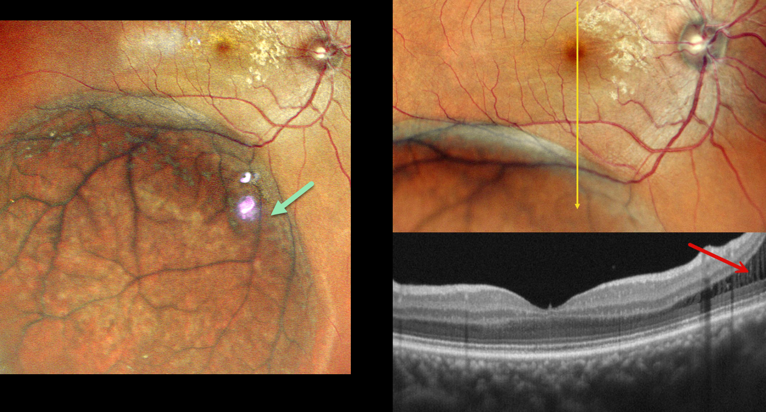
This asymptomatic 40YO female was referred for a retinal detachment in her right eye. Vision was 20/20 OD and 20/20 OS in her normal OS.
Pseudocolor SLO imaging shows a large area of inferotemporal degenerative retinoschisis (DRS) extending just into the distal macula. OCT scanning confirms schisis without retinal detachment. Observation was recommended.
Learning Points:
Uncomplicated DRS typically follows a stable or slowly evolving course that should not be treated. The development of inner-layer and outer-layer holes essentially creates a full-thickness retinal break. Progression of subretinal fluid beyond the DRS margins (combined schisis-rhegmatogenous retinal detachment, RRD), in the presence of both inner- and outer-layer holes, is the only DRS scenario requiring intervention.
It is often clinically difficult to differentiate pure DRS from a combined schisis-RRD or from RRD. OCT scanning through the area of interest, as in our patient, often provides definitive evidence for the proper diagnosis and management (Paris et al, Survey Ophthalmology 2026;71:71-80).



