

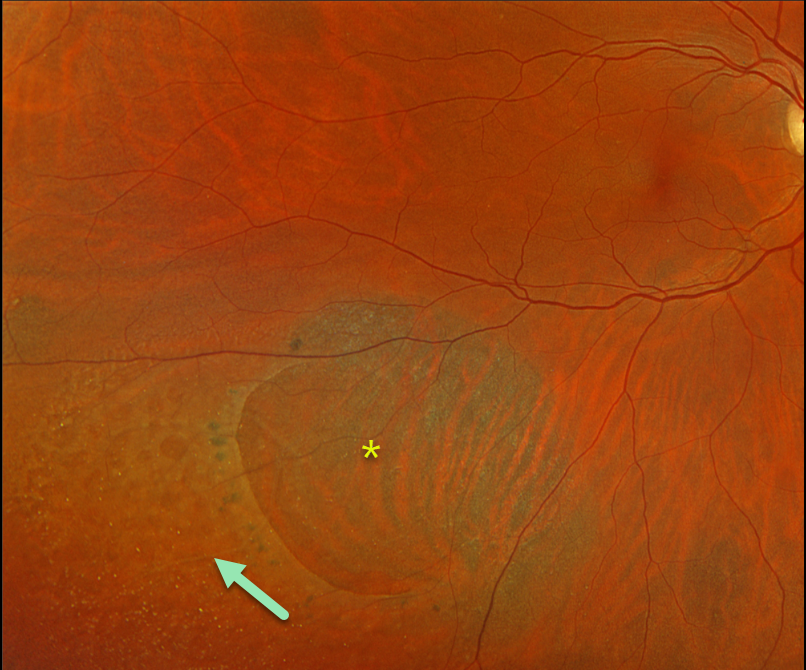
This 61YO female has been followed for several years for asymptomatic unilateral degenerative retinoschisis in her right eye. Vision is 20/30.
Optos color RGB imaging shows a large posterior outer layer hole with surrounding pigment. Innumerable small inner-layer holes are noted more peripherally within the inferotemporal schisis cavity.
Learning Points:
Degenerative retinoschisis is virtually never prophylactically treated (see Byer 1986;93:1127-1137). Patients can rarely develop a combined schisis rhegmatogenous retinal detachment (RRD) when liquid vitreous accesses the subretinal space through both an inner-layer and an outer-layer hole.
We will therefore continue to follow her twice yearly.



