

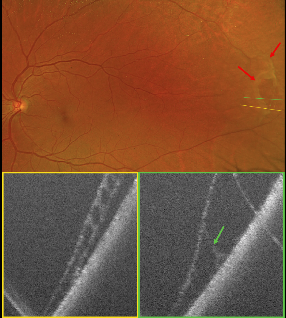
This 64YO female was referred for a possible asymptomatic rhegmatogenous retinal detachment (RRD) in her left eye. Vision was 20/20 in her normal OD and 20/20 OS.
Optos color RG imaging shows superotemporal degenerative retinoschisis with a few outer-layer holes (OLH). OCT confirms the schisis as well as the outer layer hole.
Learning Points:
Clinically, degenerative retinoschisis usually appears as a smooth, dome-shaped peripheral retinal elevation without the outer retinal corrugations classically seen in RRD (Oquendo et al, AJO 2024;268:212-221). Degenerative retinoschisis is virtually never prophylactically treated (Byer 1986;93:1127-1137). Although the schisis cavities can enlarge, prophylactic treatment, including thermal laser, does not prevent further spread and should not be performed (Ness et al, Surv Ophthalmology 2022;67:892-907).
Situs inversus of the optic disc was identified as a benign incidental finding in our patient. This occurs in 5% of normal eyes and 70% of eyes with tilted discs (Witmer et al, Surv Ophthalmology 2010;55:403-428). It can be mistaken for other congenital disc anomalies.



