

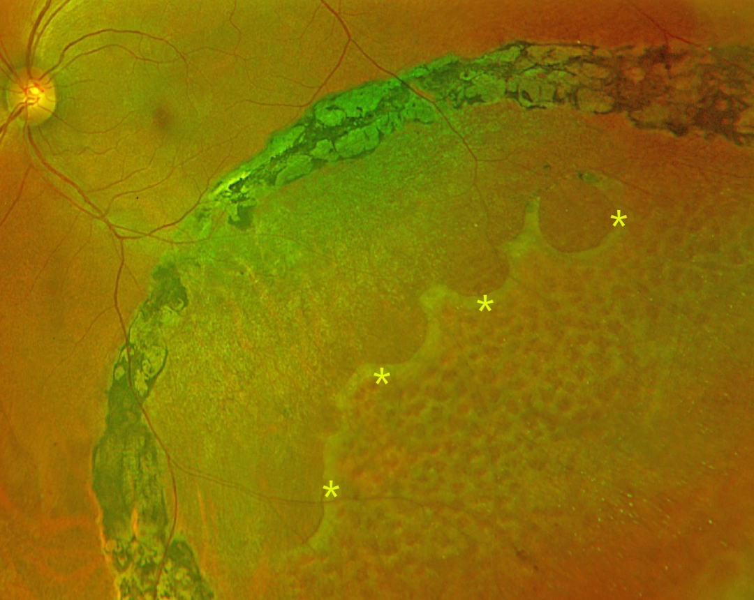
This 47YO female presented with 4 years of flashes in her left eye that began immediately following laser photocoagulation elsewhere. Vision was 20/20 bilaterally.
Optos color RG imaging shows inferotemporal degenerative retinoschisis, demarcated by heavy, confluent laser scarring along its posterior extent. A row of outer-layer holes is noted within the schisis cavity, with innumerable inner-layer holes more anteriorly.
Learning Points:
Degenerative retinoschisis is virtually never prophylactically treated (Byer 1986;93:1127-1137). Although the schisis cavities can enlarge, prophylactic treatment including thermal laser does not prevent further spread, should not be performed (Ness et al, Surv Ophthalmology 2022;67:892-907), and in our patient’s case, caused persistent symptomatic flashes. Vitrectomy surgery can successfully repair combined schisis rhegmatogenous retinal detachment (RRD) when liquid vitreous enters the subretinal space through holes in both the inner and outer layers.



