

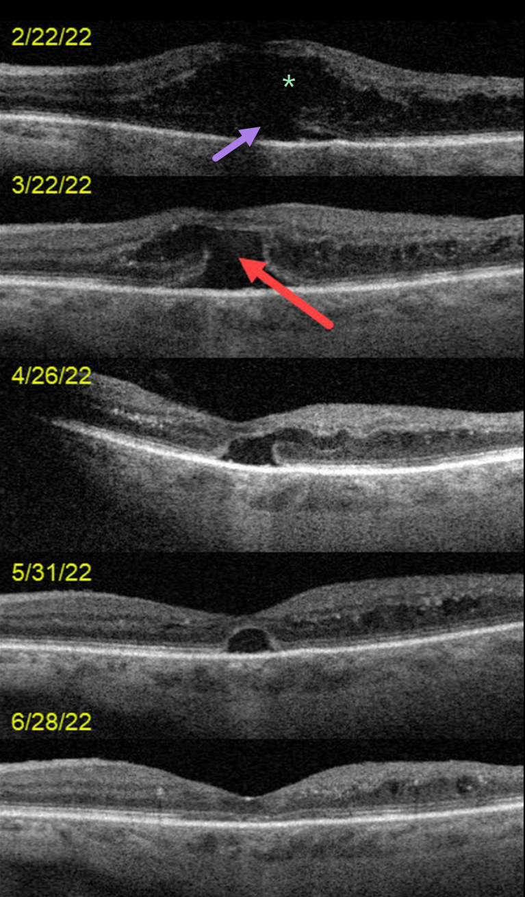
This 72YO female presented on 2/22/22 with counting-finger vision in her left eye.
OCT scanning at the initial visit shows severe center-involved diabetic macular edema (CI-DME) with shallow subretinal fluid. Anti-VEGF therapy was started.
One month later, vision remained at counting fingers. The retinal thickness had improved, but a new outer macular hole was noted.
Following 3 additional monthly injections, the edema continued to resolve with spontaneous closure of the outer macular hole. Vision was 20/70 at the 6/28/22 visit.
Learning Points:
Traditional primary full-thickness macular holes are caused by vitreomacular traction. Outer macular holes, however, have a much broader range of etiologies.
In our practice, we most commonly see these lesions in optic pit maculopathy and paraproteinemia (see Mansour et al, Ophthalmology 2014;121:1925-1932).
Our patient’s outer macular hole was likely caused by inner retinal exudation that extended through the outer retina. The return of central vision and closure of the defect, with relatively intact outer macular architecture, likely indicates that this was an outer macular dehiscence rather than a true loss of tissue. See Kumawat et al for a great review of atypical macular holes (Retina 2019;39:1236-1264).



