

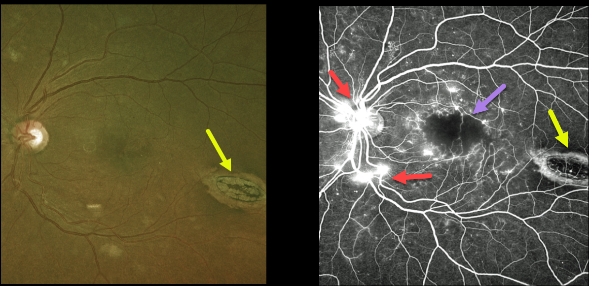
This 45YO female with type 2 diabetes presented with bilateral proliferative diabetic retinopathy (PDR). Vision in her left eye was 20/200 due to severe foveal ischemia, which is best seen on fluorescein angiography. Leaking disc and peripheral neovascularization are also seen angiographically. The macula was also edematous on OCT scanning (not shown).
An incidental finding completely unrelated to her diabetic retinopathy was an oval-shaped chorioretinal lesion in the distal inferotemporal macula known as torpedo maculopathy.
Although anti-VEGF therapy was started in her left eye for the macula edema, we doubt vision will significantly improve due to the foveal ischemia.
Learning Points:
Gass originally described torpedo maculopathy as a solitary hypopigmented nevus of the RPE (Arch Ophthalmology 1992;1358-1359). These benign, unilateral lesions are teardrop-shaped and almost always located in the horizontal meridian of the temporal macula.



