

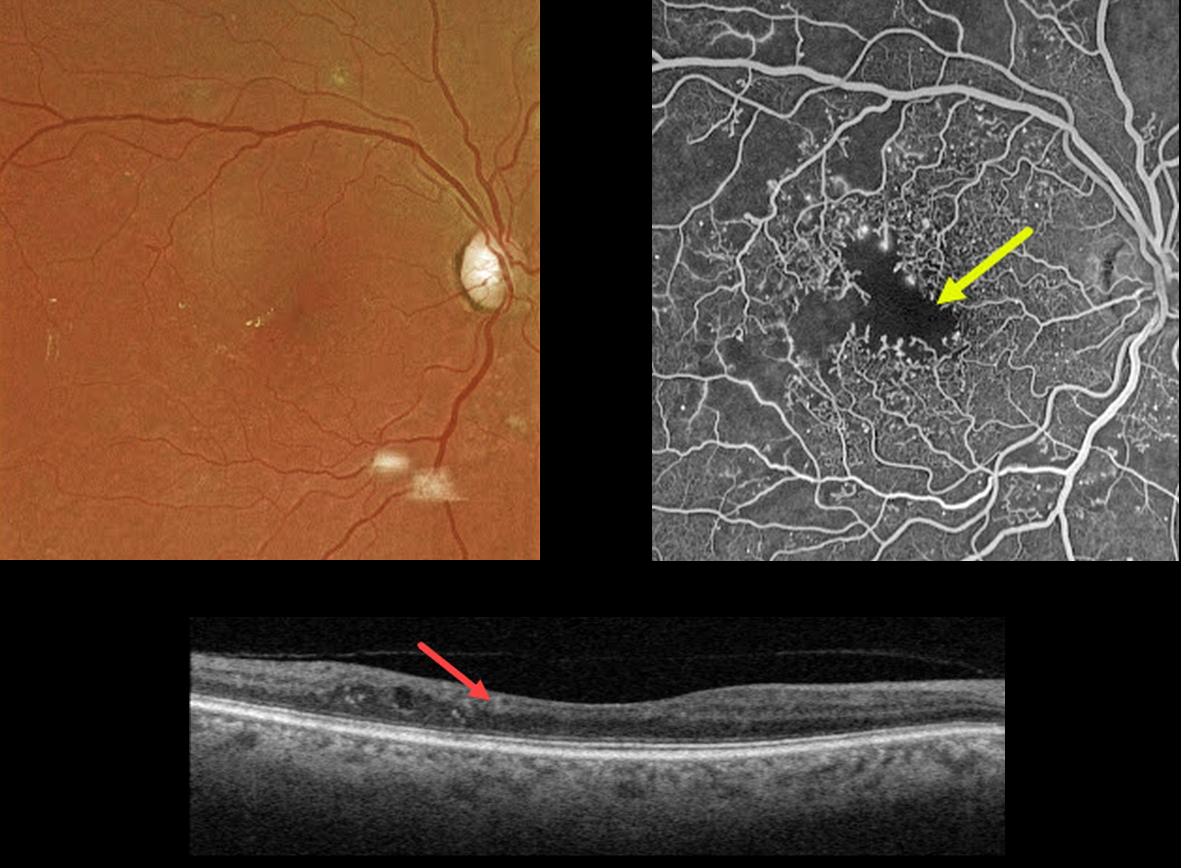
This 67YO male has a history of type 2 diabetes. Vision is 20/40.
Optos color imaging is fairly unremarkable, with some mild temporal foveal lipid. However, fundus fluorescein angiography shows foveal ischemia with an enlarged, irregular foveal avascular zone along with more peripheral superotemporal retinal ischemia.
Swept-source OCT shows a thinned, relatively featureless inner retina, also known as disorganization of inner retinal layers (DRIL).
Observation was recommended. The ischemic retina and DRIL will not improve with anti-VEGF therapy.
Learning Points:
DRIL is seen in various retinal vascular disorders, including diabetic macular edema and retinal vein occlusions. It is generally associated with worse vision and increased anti-VEGF treatment burden (see Babiuch et al, JAMA Ophthalmol 2019;137:38-46).



