

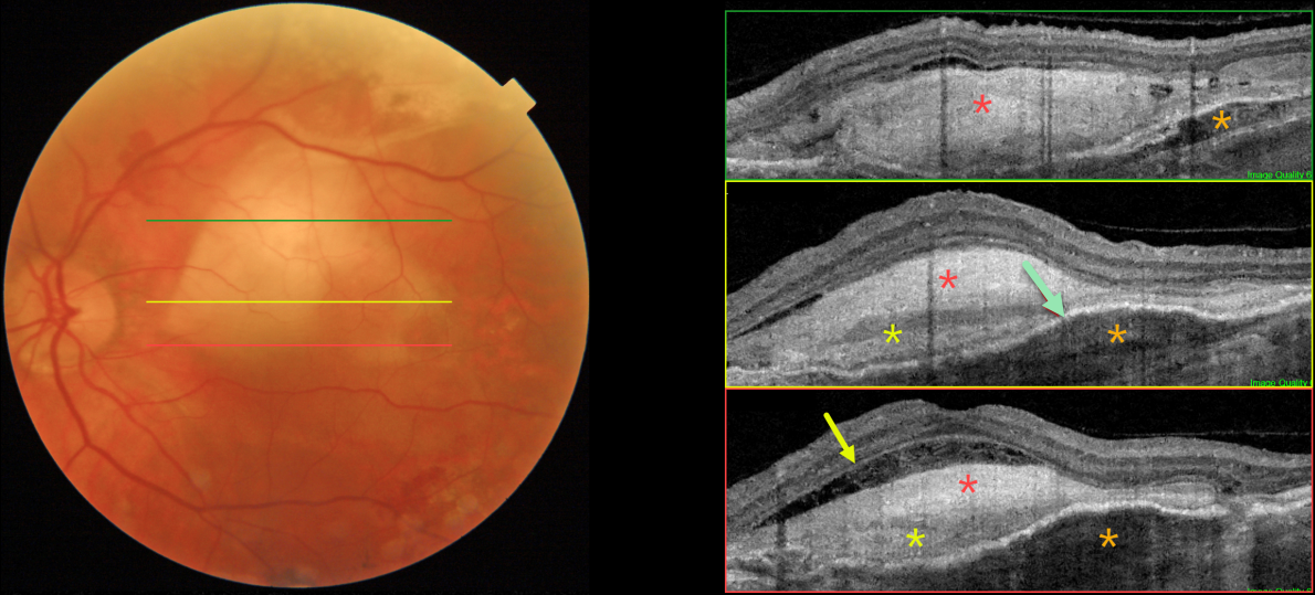
This 81YO male has a history of counting-finger vision in his left eye due to a stable age-related fibrosed disciform scar.
Color imaging shows a fibrosed disciform scar in the central and superior macula. Triton swept-source OCT through different regions of the scar shows a constellation of interesting findings. The scar is variably hyperreflective, with areas of increased and relatively decreased reflectivity. Most inferiorly, the outer plexiform layer is clearly visible overlying an area of fluid and disorganized outer retina. Superiorly, the fibrosis indents and compresses the underlying RPE and choroid. Finally, the fibrosed neovascular lesion is both above (type 2) and below (type 1) the RPE, with the RPE variably elevated.
Learning Points:
Disciform scars were the norm for untreated wet AMD prior to the anti-VEGF era that began in the mid-2000s. This type of scarring is virtually no longer seen in eyes treated early in the course of their neovascular disease, although some subretinal fibrosis remains in 40% of eyes after 10 years of anti-VEGF treatment (Chong et al, Retina 2020;40:2285-2295).



