

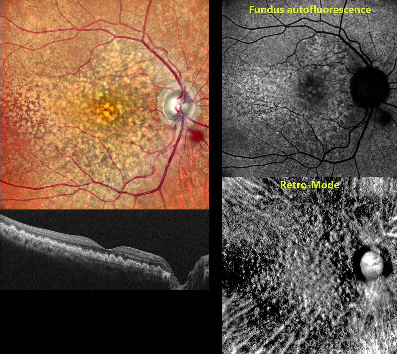
This 58 YO female presented to us with 2 to 3 years of bilateral vision loss. There was no family history of eye disease. Vision was 20/20 OU.
Pseudocolor SLO imaging shows extensive mixed and confluent drusen extending throughout the right posterior pole. OCT scanning shows drusen indenting the overlying retina, extending into the outer nuclear layer and, in some areas, up to the outer plexiform layer. Fundus autofluorescence (FAF) shows macular hyper-FAF with surrounding relative hypo-FAF. Retro-Mode imaging reveals a dramatic, almost 3-dimensional appearance of depressions and mounds resembling the surface of the moon. Identical findings were noted in her left eye (not shown).
Learning Points:
Doyne’s, also known as dominantly inherited radial basal laminar drusen or Malattia Leventinese, is a rare macular disorder caused by a mutation in the EFEMP1 gene on chromosome 2p16, although we have seen similar phenotypes with negative genetic testing. The EFEMP1 protein is a member of the fibulin family of extracellular matrix glycoproteins. The defective protein creates an abnormally thickened RPE basement membrane. Centrally large, nodular, and confluent drusen develop, along with a temporal radiating pattern of smaller cuticular drusen. Later, there may be variable amounts of RPE hyperplasia and fibrous metaplasia. Macular neovascularization may also occur.
Fundus autofluorescence (FAF) usually shows hypo-FAF in age-related drusen. The lesions in Doyne’s, as in this patient, usually show hyper-FAF, likely from unmasking of the RPE by overlying outer retinal thinning.



