

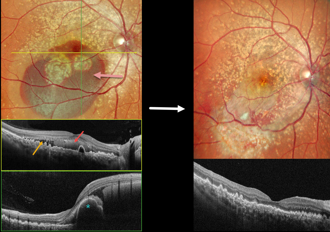
This 49YO female presented with 3 days of decreased vision in her OD. Vision was 20/70 OD and 20/15 OS. There was no family history of eye disease.
Pseudocolor SLO imaging shows extensive mixed and confluent drusen extending through each macula into the midperipheries. Inferior submacular blood extends into the right foveal center. OCT scanning shows extensive conically shaped drusen. The subretinal blood in her right eye is hyperreflective, with additional temporal subretinal fluid. A bilobed nodular RPE detachment is present within the nasal blood.
She underwent pars plana vitrectomy, fluid-air exchange, and intravitreal Avastin. One month postoperatively, vision improved to 20/15 OD with resolved fluid and trace residual blood.
Learning Points:
Doyne’s honeycomb macular dystrophy, also known as dominantly inherited radial basal laminar drusen or Malattia Leventinese, is a rare macular disorder caused by a mutation in the EFEMP1 gene on chromosome 2p16. The EFEMP1 protein is a member of the fibulin family of extracellular matrix glycoproteins. The defective protein creates an abnormally thickened RPE basement membrane.
Centrally large, nodular, and confluent drusen are noted, along with a temporal radiating pattern of smaller cuticular drusen. Later, there may be variable amounts of RPE hyperplasia and fibrous metaplasia. Macular neovascularization, as in this case, may also develop. Our patient’s pre-op right OCT was suggestive of polypoidal choroidal vasculopathy, although this is not a known association with Doyne’s.



