

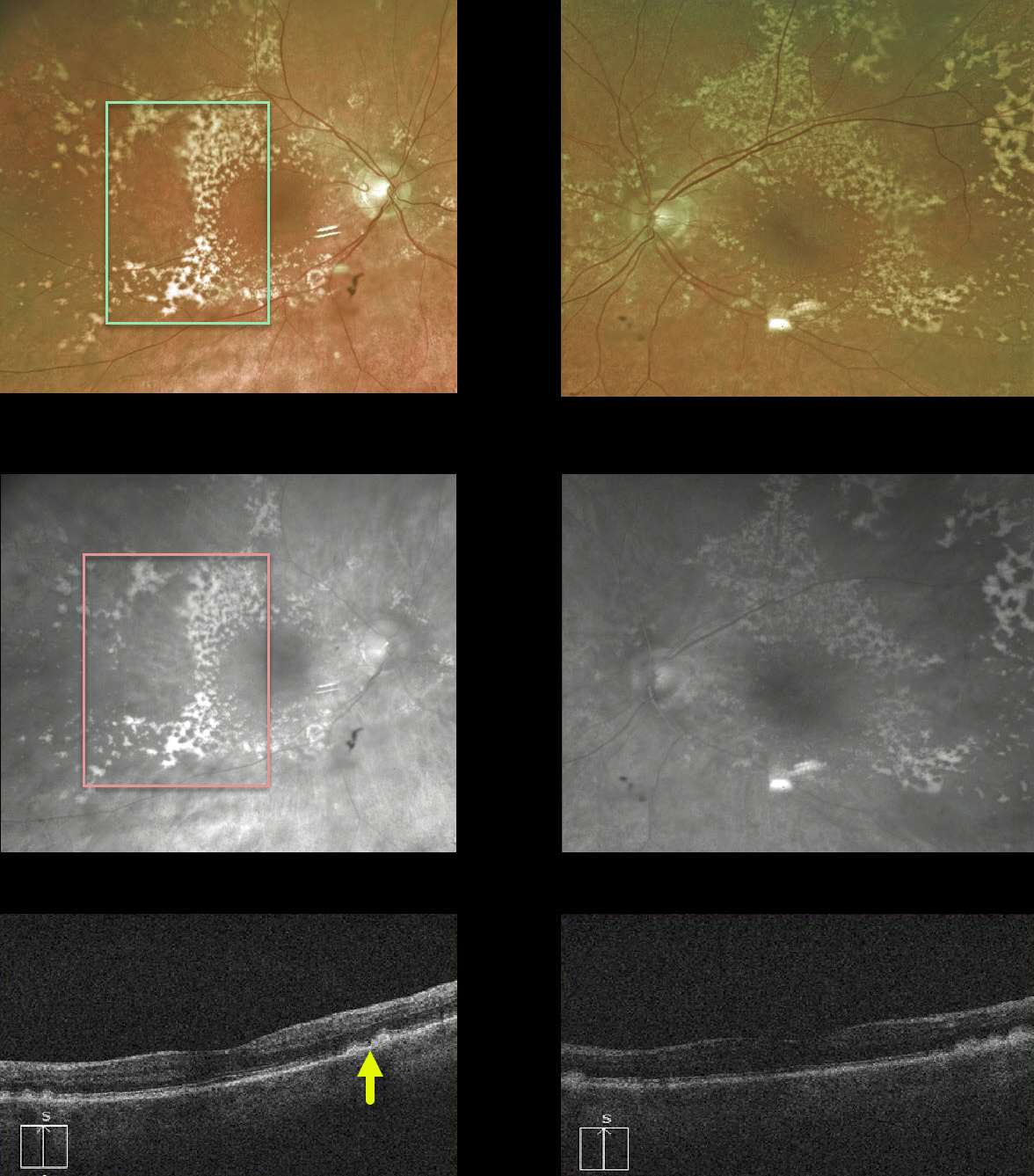
This 83YO female has extensive, confluent drusen outside the perifoveal area.
The drusen, located beneath the RPE, are well visualized with the Optos red channel. Subretinal drusenoid deposits (not present), which form above the RPE, would disappear with the red channel.
OCT also demonstrates drusen.
Learning Points:
Sub-RPE drusen are a hallmark feature of non-exudative age-related macular degeneration.



