

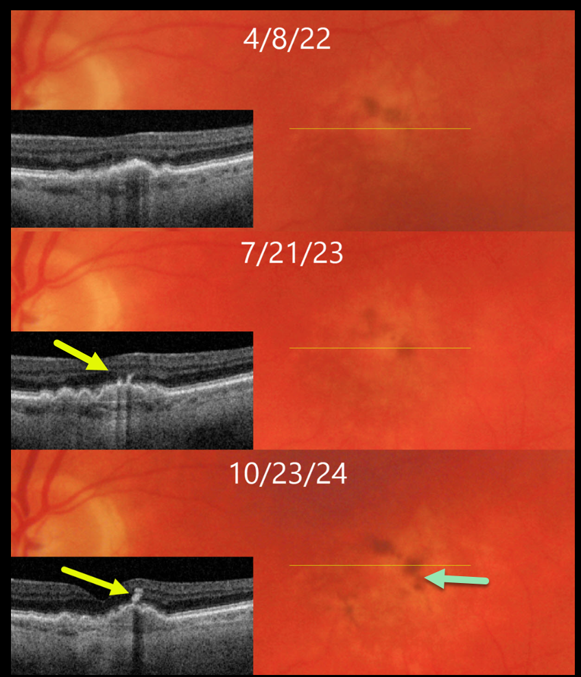
This 71YO male has been followed for several years with intermediate AMD.
On 4/8/22, vision was 20/40 in his asymptomatic left eye. There is a shallow RPE detachment (PED) with faint surrounding drusen and pigment loss on both Triton color imaging and swept-source OCT. Clumping of subretinal pigment is noted along the superior edge of the PED on color imaging.
He returned for his 1-year examination on 7/21/23 with stable 20/40 vision. A new pigment clump is noted along the temporal edge of the PED, and this is seen as a hyperreflective plume on OCT.
On 10/23/24, vision decreased to 20/100, with extension of the plume into the inner retina. There is continued remodeling of the pigment clumps on color imaging.
Learning Points:
The widely used Beckman AMD clinical classification defines intermediate AMD as including large drusen > 125 microns and/or any AMD pigmentary abnormalities (Ferris et al, Ophthalmology 2013;120:844-851). This classification was modified in 2024 to include a third risk factor, subretinal drusenoid deposits, which significantly increases the risk for progression to late AMD (Agron et al, Ophthalmology 2024;131:1164-1174).
These pigment clumps, which are visualized on OCT as hyperreflective foci (HRF), are found in over 50% of eyes with AMD (Ho et al, Ophthalmology 2011;118:687-693), and are thought to represent thickened RPE or intraretinal migrating RPE cells (Laiginhas et al, Ophthalmology Science 2022;2:100116). The migrating RPE cells often track along Muller cells creating a hyperreflective plume (Cao et al, IOVS 1021;62(10):34). These RPE cells transdifferentiate into macrophage-like cells. Macular atrophy will often develop underlying the HRF.



