

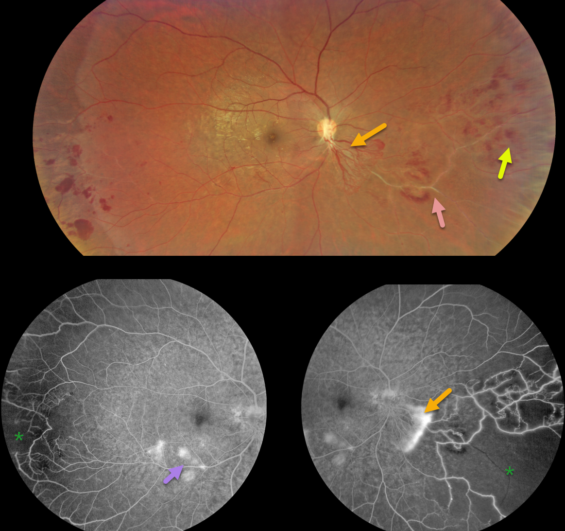
This healthy 31YO male presented with 6 months of blurred vision. He was previously diagnosed by a general ophthalmologist with a branch vein occlusion in the OD. Vision was 20/30 in his right eye and 20/20 in his left eye.
Color imaging shows active neovascularization extending inferonasally from the nerve, with more distal retinal hemorrhages and vascular sheathing.
Fluorescein angiography shows leaking neovascularization with retinal ischemia nasally and temporally. Some vascular leakage is noted in the inferotemporal macula.
Learning Points:
First described by British ophthalmologist Henry Eales in 1880, Eales disease is characterized by sharply circumscribed peripheral regions of distal ischemia, often with retinal neovascularization at the junction of the perfused and ischemic retina. It is more common in males in their 20’s to 30’s and may be associated with a prior exposure to tuberculosis. Most patients are asymptomatic unless they develop a vitreous hemorrhage. Scatter laser to ischemic retina and intravitreal anti-VEGF injections are effective for retinal neovascularization.
It is likely a heterogeneous disorder and is a diagnosis of exclusion, needing first to rule out the multitude of other disorders that can cause peripheral retinal ischemia and neovascularization. The location of our patient’s neovascularization is a bit unusual for Eales, which is usually more peripheral. However, since his systemic work-up, including chest X-ray, was normal except for a positive PPD, we feel Eales is a reasonable diagnosis. A scatter laser to the ischemic retina has been planned and scheduled.



