

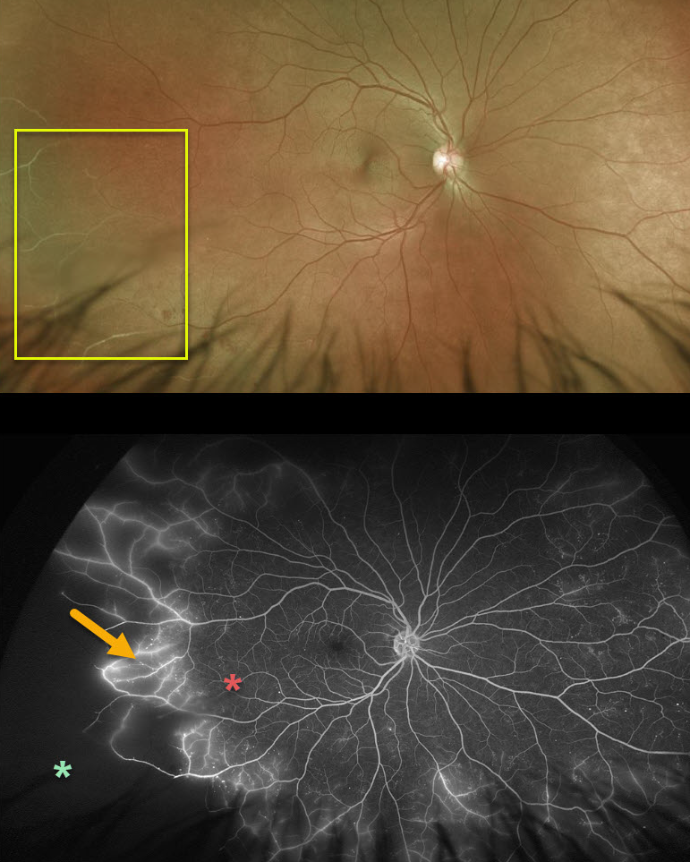
Eale’s disease is our best guess for this 44YO male patient. Eale’s disease is characterized by peripheral, sharply circumscribed regions of ischemia. Fluorescein angiogram demonstrates leakage of retinal vessels at the junction of perfused and ischemic retina.
For now, we are carefully observing the ischemic retina, but we will treat this region with scatter laser if neovascularization develops.
Learning Points:
Eale’s disease is an idiopathic peripheral occlusive retinal vasculopathy that can later develop peripheral neovascularization. It is more common in males in their 20’s to 30’s, and may be associated with prior tuberculosis exposure. Most patients are asymptomatic unless they develop a vitreous hemorrhage.
It is likely a heterogeneous disorder and is a diagnosis of exclusion, needing first to rule out the multitude of other disorders that can cause peripheral retinal ischemia and neovascularization.
We have seen many patients with these findings since integrating Optos ultrawidefield imaging into our practice, and therefore suspect that these peripheral retinal findings are much more common than previously described.
Peripheral retinal microvascular abnormalities have also recently been described with ultrawidefield imaging, and may represent a normal variant in this retinal vascular watershed zone (see Shah et al, Retina 2016;36;1087-1092).



