

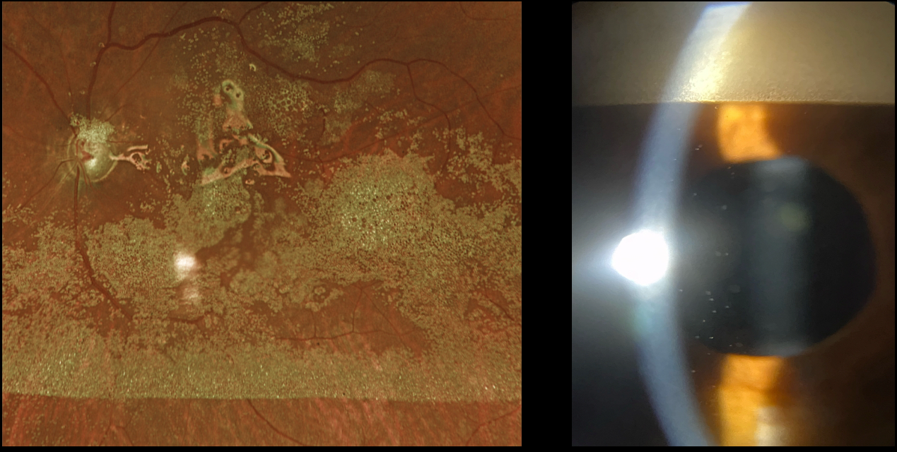
This 65YO male underwent vitrectomy surgery with silicone oil 5 years earlier for a complex rhegmatogenous retinal detachment with proliferative vitreoretinopathy. Vision has remained at counting fingers since the surgical repair, and he presented for his annual visit without new complaints.
Optos RG imaging shows innumerable tiny droplets of emulsified silicone oil coating the posterior pole. Slit lamp photography shows a reverse ‘hypopyon’ of emulsified oil superiorly. Suspended oil droplets are also noted throughout the anterior chamber.
Learning Points:
Silicone oil as an adjunct for repairing complex retinal detachments was introduced by Paul Cibis in the early 1960s (Cibis et al, Arch Ophthalmol 1962;68:590-599). It is used primarily for complex detachments at high risk for failure, including severe proliferative vitreoretinopathy and viral retinitis.
Although silicone oil is generally well-tolerated as a long-term tamponade, it can occasionally emulsify, resulting in numerous tiny, opaque droplets. Emulsification is multifactorial, involving the properties of the oil, the surgical procedure, and postoperative factors.
When severe, emulsified droplets can cause significant vision loss and require removal. The droplets can also migrate into the anterior chamber, causing a reverse ‘hypopyon’ with secondary elevated intraocular pressure or corneal decompensation.
Our patient’s increased intraocular pressure of 32 mmHg, which was elevated for the first time, was attributed to emulsified oil clogging the trabecular meshwork. Topical glaucoma drops were started. If the pressure cannot be adequately controlled medically, then vitrectomy with removal of the emulsified oil and likely exchange for new silicone oil will be recommended.



