

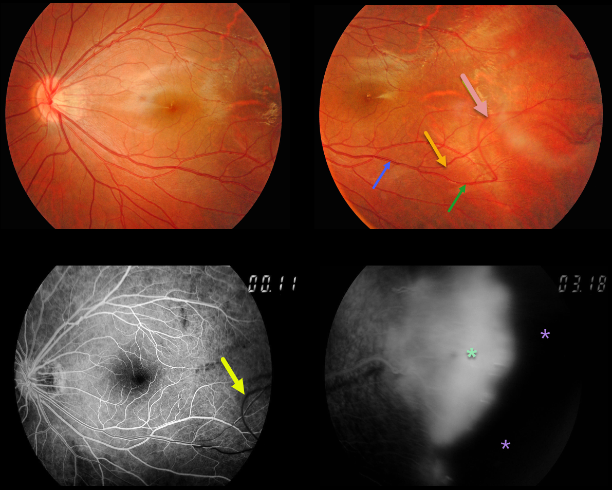
Our patient’s macular retinal vessels are dragged temporally. His brother has similar retinal findings.
The major inferotemporal vein branches into smaller superior and inferior venules. The inferior venule makes an abrupt turn more distally, arching superiorly before extending into the temporal periphery.
Arteries cross veins, veins cross arteries, but arteries never cross arteries, and veins never cross veins.
So why does this inferior venule eventually cross the superior venule? Although we don’t have any stereoscopic images, we suspect that vitreous traction pulled the inferior venule anteriorly and superiorly.
This is also evident on fluorescein angiography (FA), where the superior vessel, which has still not completely filled, is clearly anterior to the other venules, which have laminar filling. FA also shows leaking neovascularization and more peripheral retinal ischemia.
Learning Points:
Originally described by Criswick and Schepens (AJO 1969;58;578-594), familial exudative vitreoretinopathy (FEVR) can be inherited as an autosomal-dominant, recessive, or X-linked trait with high penetrance and variable expressivity.
FEVR is characterized by peripheral temporal retinal avascularity, lipid exudation, neovascularization, tractional retinal detachment, and temporal dragging of the macula and retinal vessels. These findings are somewhat similar to those found with retinopathy of prematurity (ROP).



