

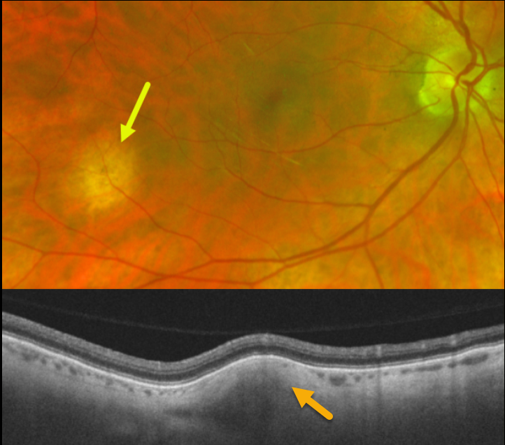
Optos color imaging shows an amelanotic subretinal lesion in the right inferotemporal midperiphery. Swept-source OCT through this lesion shows hyperreflective scleral thickening, which compresses the overlying choroid.
Learning Points:
Focal scleral nodule (previously known as solitary idiopathic choroiditis [SIC] and unifocal helioid choroiditis), presents as a single, yellow-white, elevated nodular, avascular subretinal lesion. On OCT it arises from the inner sclera, causing marked choroidal compression.
For the largest series to date and the rationale behind the renaming of this entity, see Fung et al, Ophthalmology 2020;127:1567-1577.



