

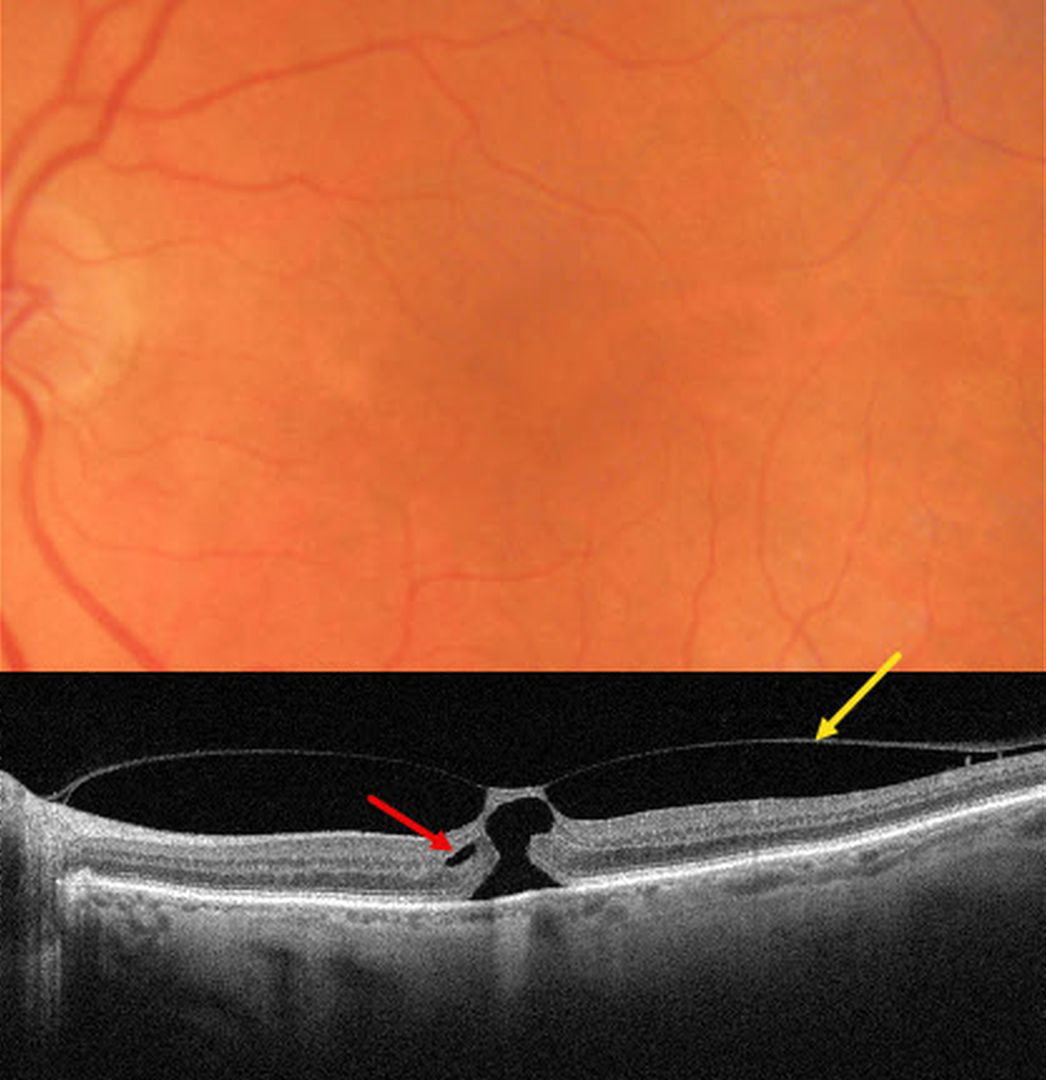
This 82YO female was referred for an asymptomatic macular finding noted on screening OCT scanning. Vision was 20/200.
Triton color imaging shows a poorly defined foveal reflex. Swept-source OCT shows a partial macular posterior vitreous detachment (PVD) that is adherent to the foveal center. A thin strip of intact inner tissue is adherent to the overlying vitreous with an underlying outer macular hole. A tiny area of split outer plexiform layer is noted nasally.
Learning Points:
The International Vitreomacular Traction Study Group classified vitreomacular traction by size (focal <= 1500 microns or broad >1500 microns) and etiology (isolated or concurrent, Ophthalmology 2013;120:2611-2619). The prevalence of vitreomacular traction (VMT) increases with age and is present in about 1% of people over 40YO. The natural history is still being defined, but most patients have stable traction and vision. VMT can also resolve spontaneously in about 20% (Errera et al, Ophthalmology 2018;125:701-707).
Management includes observation, pneumatic vitreolysis, or vitrectomy. In our experience, most patients with VMT remain completely asymptomatic despite impressive OCT findings. We therefore almost always observe VMT unless the patient has significant symptoms. Our patient elected to continue observation since she was asymptomatic bilaterally.



