

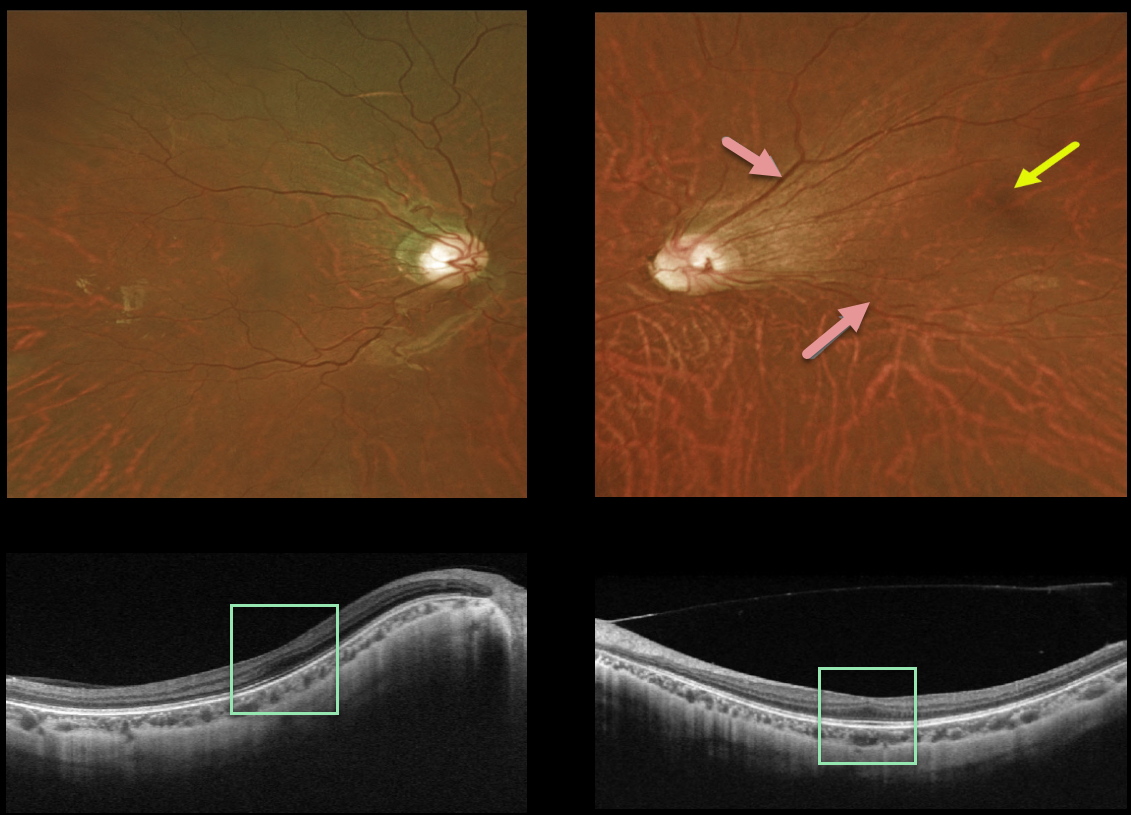
This 20YO female presented with these asymptomatic macular findings. She was born prematurely at 24 weeks of gestation. Vision was 20/25 OU.
The right posterior pole was fairly normal clinically, although the left major temporal arcades were straightened and dragged temporally. The left fovea was also temporally displaced.
Triton swept-source OCT showed a flattening of the normal foveal depression bilaterally.
Learning Points:
Temporal dragging and straightening of the vascular arcades are common findings in regressed retinopathy of prematurity (ROP). This is caused by involuted peripheral temporal neovascularization displacing the posterior retina.
The macular center is usually about 2 disc diameters temporal to the edge of the optic nerve, and our patient’s is about 3 disc diameters.
Fovea plana is characterized by the absence of the foveal pit, which is formed embryologically by the inner retinal tissues and vasculature being displaced centrifugally.
It is most commonly associated with albinism, prematurity (as in our case), or as an isolated finding. Despite the abnormal foveal architecture, vision is usually normal.



