

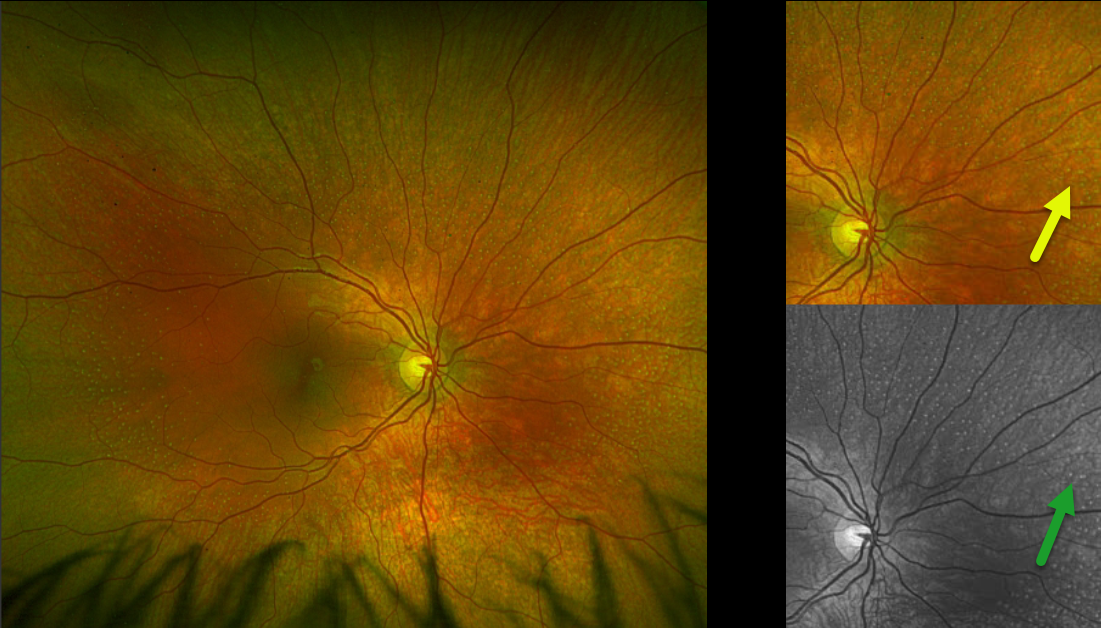
This healthy 10YO girl was referred for asymptomatic findings on retinal examination. There was no ocular family history. Vision was 20/40 OU.
Optos color RG imaging of the right eye shows innumerable subretinal yellow-white dots extending from outside each macula into the midperiphery. These dots are most evident in green-channel imaging. Identical findings were noted in the left eye (not shown). Genetic testing revealed double heterozygous RDH5 mutations.
Learning Points:
Fundus albipunctatus, an autosomal recessive form of congenital stationary night blindness, presents with scattered yellow-white dots that extend into the midperipheries, sparing the macula. Patients experience stable night blindness with normal central acuity. It is caused by a mutation in the RDH gene, which encodes the retinol dehydrogenase protein. This protein, found in the RPE helps recycle vitamin A in the visual cycle, and the white dots are thought to contain 11-cis retinal precursors. This results in decreased retinoid-derived fluorophores, accounting for the diminished FAF in these eyes (Sergouniotis et al, Ophthalmology 2011;118:1161-1670).



