

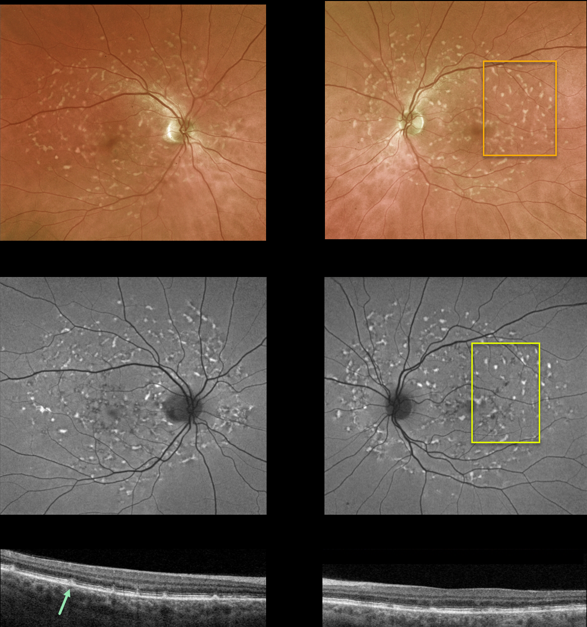
This 55YO female was referred for a possible white dot syndrome. She had no ocular symptoms, and vision was 20/30/ OU. There was no family history of ocular disease.
Optos color imaging shows multiple yellow fleck-like lesions throughout each posterior pole. Fundus autofluorescence of these lesions shows variable hyper-FAF.
OCT scanning shows scattered defects in the EZ with hyperreflective dots extending from the RPE into the EZ and outer nuclear layer.
Learning Points:
Fundus flavimaculatus is a genetic disorder, usually transmitted as an autosomal recessive trait, that is caused by mutations involving the ABCA4 gene. This gene encodes one of a family of ATP-binding cassette (ABC) transmembrane proteins, which mediate the active transport of various substrates across cellular membranes. The ABCA4 protein is present in the photoreceptor outer segment disc membranes, where it participates in the recycling of 11-cis-retinal.
Stargardt disease is believed to be fundus flavimaculatus without peripheral findings. Vision in fundus flavimaculatus is often better than that in Stargardt due to later disease onset and less macular involvement. Both likely represent an RPE lipofuscin storage disease. The RPE cells become engorged with lipofuscin, causing a dark choroid on fluorescein angiography.
See Cremers et al (Progress Retinal Eye Research 2020;79;100861) for a wonderful review of the complex spectrum of ABCA4 disorders.



