

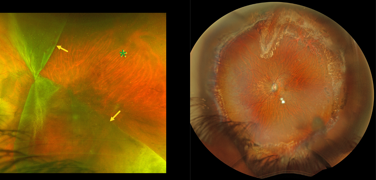
This 44YO male presented with hand motion vision OS from an acute giant tear (GRT) retinal detachment. Optos color RG imaging shows a complex GRT with one flap folded superonasally and the other inferotemporally, creating a sharply defined square region of temporal bare RPE. The optic disc is covered by the giant tear. Emergent vitrectomy was scheduled.
Six months postoperatively, the retina remained completely attached. Vision was 20/80.



