

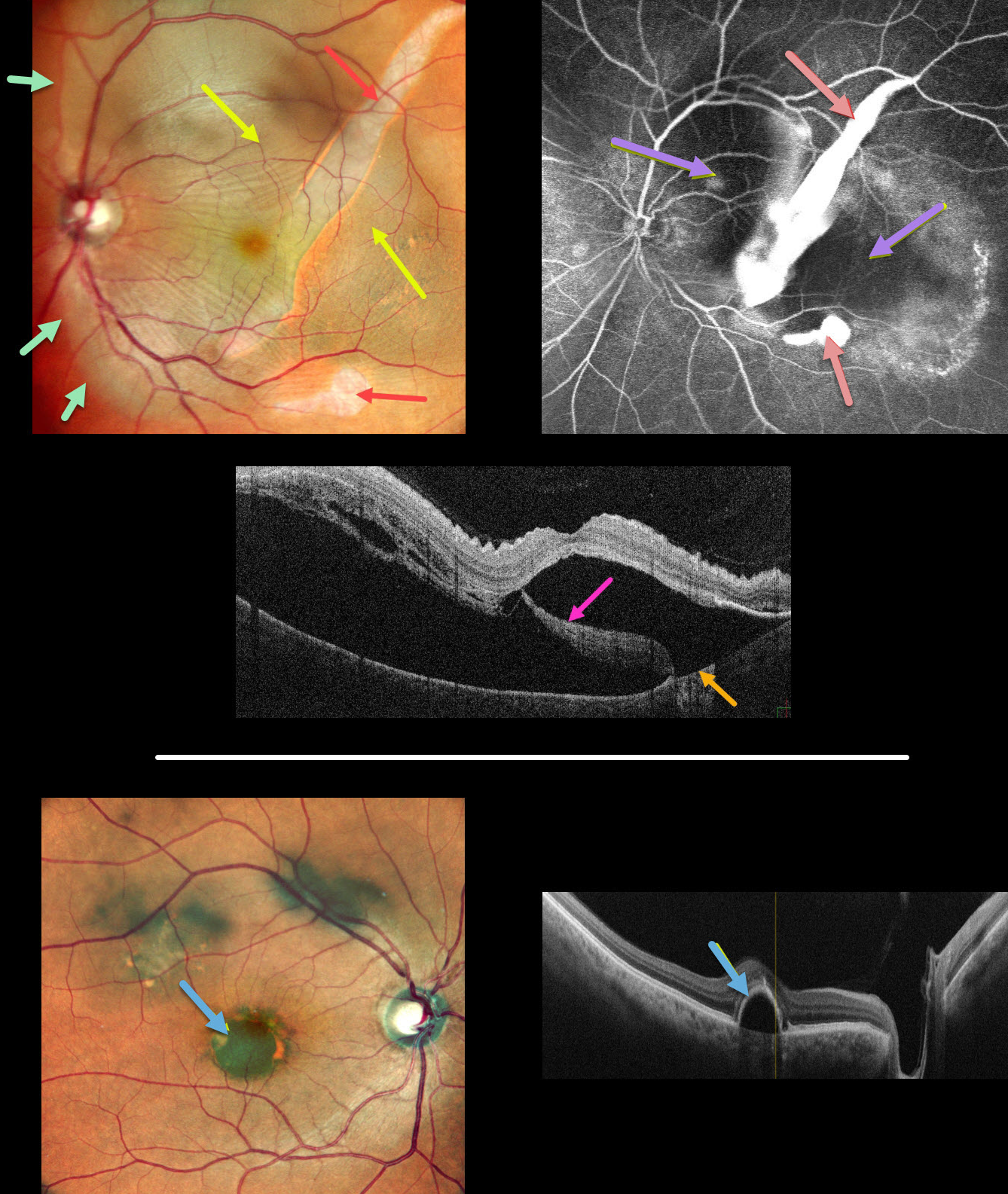
This 62YO male presented with 1 month of vision loss in his left eye. Vision was 20/30 OD and 20/60 OS.
Multicolor imaging of his left eye shows a giant RPE tear extending from the inferior macula into the superotemporal midperiphery, along with a much smaller tear just inferior to the macula. The beds of the RPE tears are depigmented. Increased subretinal pigmentation is noted on either edge of the giant tear. Subretinal fluid variably elevates the macula with inner retinal folds radiating from the optic nerve temporally. This subretinal fluid extends into the superior and temporal midperipheries.
B-scan OCT shows marked subretinal fluid. Bare Bruch’s membrane is noted temporally, and the RPE tear is lifted nasally and floating within the serous detachment.
Fluorescein angiography shows marked hyperfluorescent window defects from the absent RPE with blockage on either side of the giant tear from the redundant retracted RPE. He was placed on a 5-day tapering course of oral steroids but was subsequently lost to follow-up.
An asymptomatic retinal pigment epithelial detachment (PED) is noted in his right macula. The choroid does not appear thickened on OCT B-scan.
Learning Points:
A tear of the RPE is most commonly seen in RPE detachments (PEDs) associated with neovascular AMD, but can also occur in numerous other etiologies, including central serous chorioretinopathy and polypoidal choroidal vasculopathy. The tear leaves a depigmented region where the RPE was and is now absent, along with a pigmented, relatively straight subretinal lesion from the scrolled RPE edge.
Our patient’s presentation is unique in that RPE tears are usually much smaller and tend to rip in just one direction. The edges of our patient’s RPE retracted both nasally and temporally. RPE tears are usually singular, and ours had an additional, much smaller secondary tear more inferiorly.
Although our patient’s findings seem to be in the pachychoroid spectrum, the lack of a thickened choroid on OCT scanning makes the etiology less certain.
See Ersoz et al for a great review on RPE tear classification, pathogenesis, predictors and management (Surv Ophthalmology 2017;62:493-505).



