

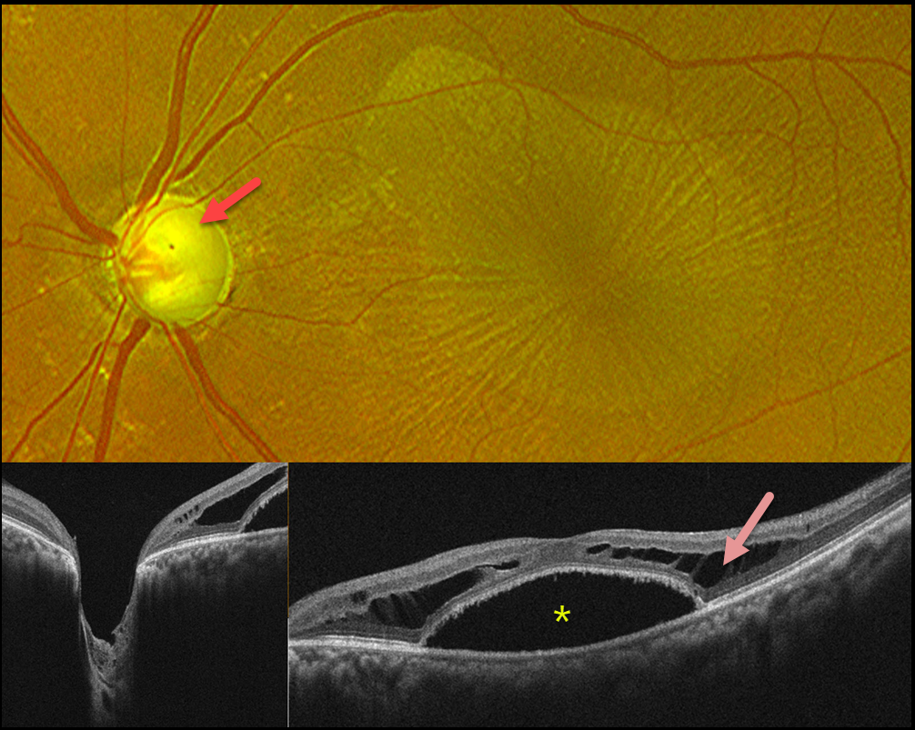
This 45YO male presented with blurred vision in his left eye. The right eye was phthisical due to absolute glaucoma. He was taking topical dorzolamide and travoprost OU. Vision was 20/400 OS. Intraocular pressure was 12 mm Hg.
Optos color RG imaging shows a nearly completely cupped glaucomatous nerve. There is an irregular oval discoloration of the outer macula with overlying radiating retinal striae. OCT scanning shows Henle’s layer schisis with a central macular detachment. An optic pit could not be identified clinically or on OCT.
Learning Points:
While peripapillary retinoschisis (PPRS) secondary to congenital optic disc cavitary abnormalities is a well-known phenomenon, similar findings in patients with glaucomatous cupping but without nerve head cavitation have more recently been described and are less common (Fujimoto et al, Ophthalmology Retina 2023;7:811-818). The retinal nerve fiber layer is most commonly involved in glaucoma, whereas in optic pit maculopathy, outer and inner nuclear layer schisis, foveal involvement, and subretinal fluid are more frequently noted.
Most cases of glaucoma-related PPRS are asymptomatic and spontaneously resolve. However, in fovea-involving or symptomatic cases, surgical intervention may improve vision and decrease time to resolution. There is no consensus regarding the optimal treatment approach, including whether to perform pars plana vitrectomy alone or with the addition of juxtapapillary laser treatment, ILM peeling, inner retinal fenestration, and/or autologous platelet concentrate.
We recommended adding brimonidine and timolol drops to further lower his IOP. We are not planning any surgical intervention.



