

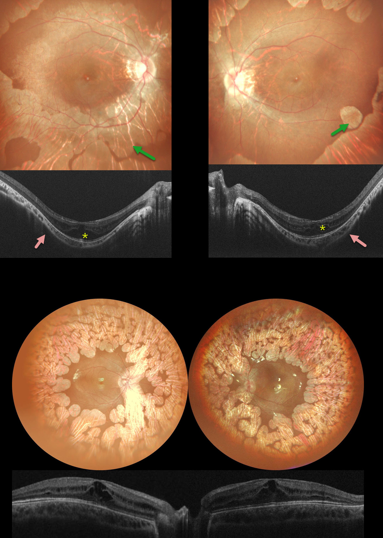
This 6YO girl has a history of night blindness, and her twin brother has a history of milestones delay and seizures. Family history was suggestive of consanguinity. The girl’s vision was 20/120 OD and 20/80 OS, and 20/120 OU in the boy.
The girl’s fundus photography shows variably coalescent, scalloped areas of bilateral chorioretinal atrophy extending from the peripheral maculas into each periphery. OCT shows bilateral cystic retinal edema primarily in the inner and outer nuclear layers. There are shallow poster staphylomas. Her brother shows similar findings, but without the staphylomas.
Blood tests of both siblings revealed a deficiency of the ornithine aminotransferase (OAT) enzyme. Topical non-steroidal anti- inflammatory eye drops were started along with an arginine-free diet and B6 supplementation.
Learning Points:
Gyrate atrophy is a rare autosomal recessive retinal dystrophy, characterized by extensive areas of lobular peripheral chorioretinal degeneration that gradually spread posteriorly. Macular involvement on OCT is common, with foveal thickening in younger patients followed by atrophy with outer retinal tubulation later in life (Sergouniotis et al, Ophthalmology 2012;119:596-605).
End-stage disease can be indistinguishable from choroideremia. Other findings include myopia and night blindness. The degeneration is caused by a lack of a vitamin B6-dependent ornithine aminotransferase mitochondrial enzyme, which leads to elevated ornithine levels. Vitamin B6 supplementation and an arginine-restricted diet



