

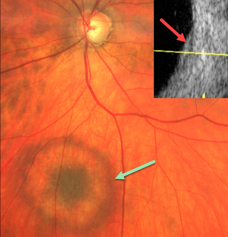
During a routine glaucoma examination, this 61YO male was found to have a halo choroidal nevus in his right eye. The nevus was hyperpigmented centrally, then encircled by relative depigmentation, and finally surrounded by a hyperpigmented rim. The lesion was clinically minimally thickened and measured about 2-3 disc diameters. The nevus was barely visible with B-scan ultrasonography.
Learning Points:
Halo choroidal nevi are a relatively rare variant of the more typical diffusely hyperpigmented lesions. A large retrospective series by Shields et al found that these lesions may be associated with cutaneous melanoma (Arch Ophthalmology 2010;128:859-864).
Malignant transformation to uveal melanoma is reduced when a halo is present (Shields et al, Arch Ophthalmol 2009;127:981-987), although this does not preclude malignant transformation.



