

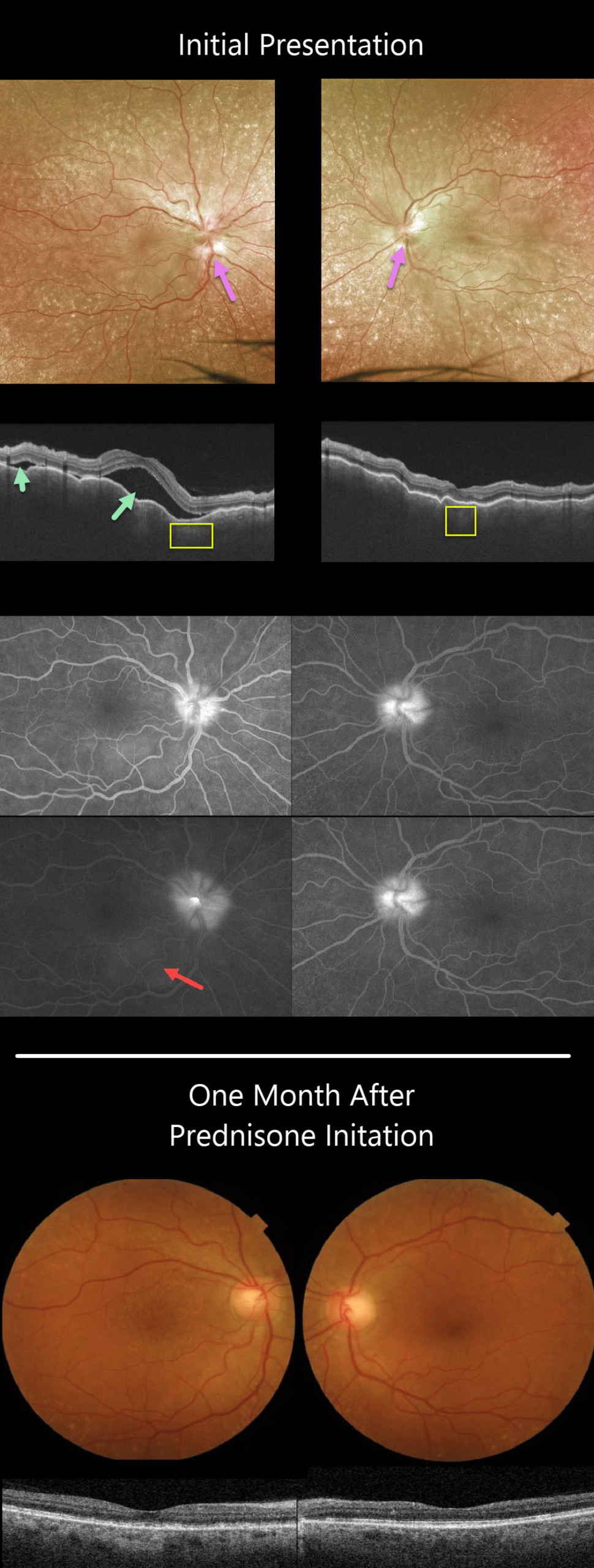
This 63YO female presented with a 3-week history of bilateral painful vision loss. Vision was 20/400 OD and 20/100 OS.
She had bilateral granulomatous anterior uveitis with a clear vitreous OU. Both nerves were swollen, and irregular chorioretinal folds were noted throughout each macula. Extensive incidental peripheral drusen were also present.
Triton swept-source OCT scanning shows a markedly irregularly thickened choroid OU with multifocal serous detachments in the right eye.
Optos fluorescein angiography (FA) shows bilateral optic nerve leakage and some faint subretinal leakage in the inferonasal right macula.
Oral prednisone was started, and there was near immediate improvement. One month later, vision improved to 20/40 OD and 20/60 OS, and all findings virtually normalized.
Learning Points:
This case has many classic findings for Harada’s disease, including bilateral panuveitis, optic nerve swelling, thickened choroid, chorioretinal folds, and multifocal serous retinal detachments.
We were surprised that the FA showed minimal subretinal leakage, since angiography typically shows extensive multifocal subretinal leaks with pooling in the overlying serous detachments and bacillary cavities.
Patients with just ocular findings have Harada’s disease, whereas those with additional systemic findings (including vitiligo, poliosis, headache, vertigo, and hearing loss) have Vogt-Koyanagi-Harada disease.



