

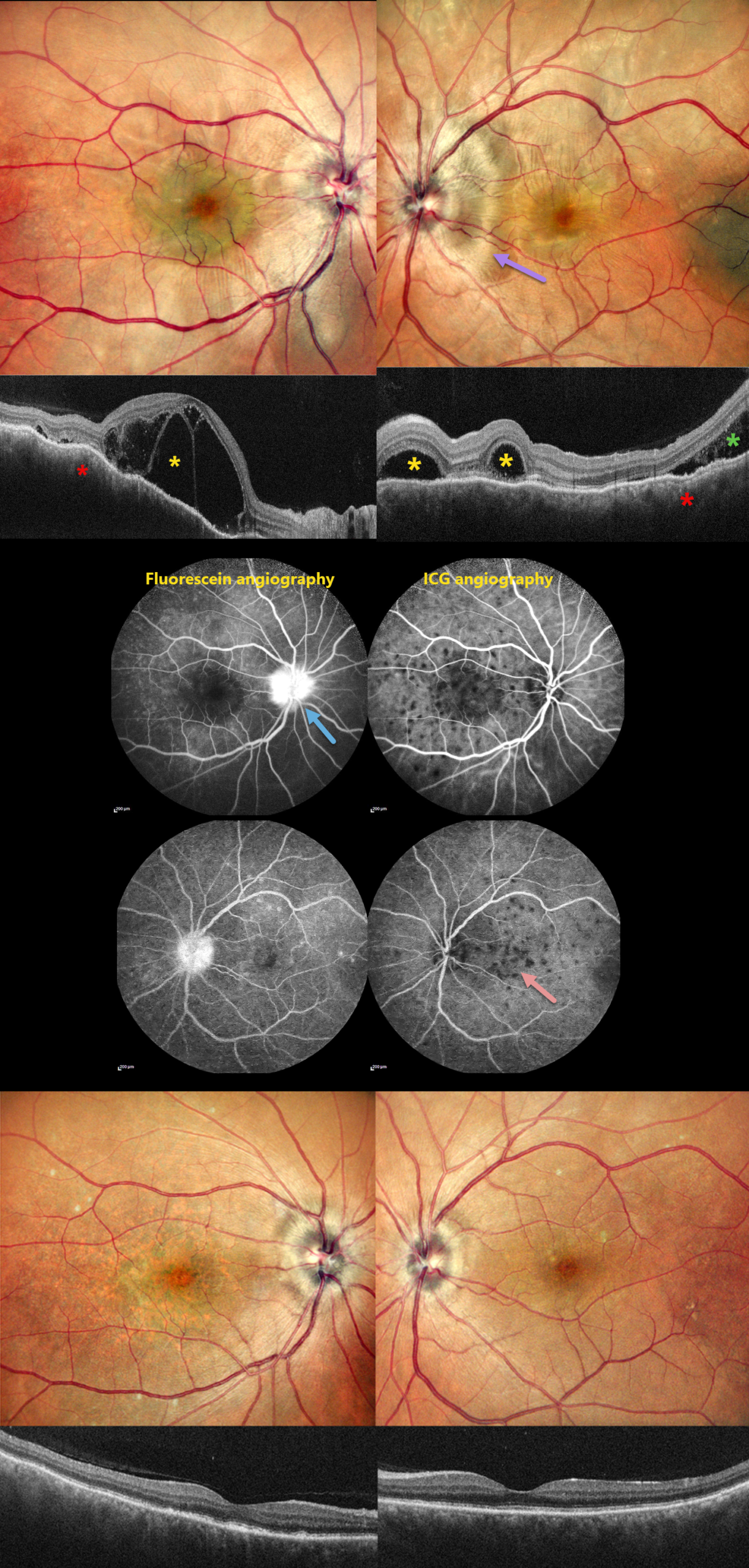
This 46YO male presented with 10 days of bilateral blurred vision with a left-sided headache. Vision was 20/200 OD and 20/20 OS. Anterior segments were normal.
Pseudocolor SLO imaging shows bilateral swollen nerves and irregular macular chorioretinal folds. OCT scanning shows multifocal bacillary layer detachments, subretinal fluid, and an undulating, thickened choroid. Fluorescein angiography shows bilateral multifocal areas of subretinal leakage and disc leakage, and indocyanine green angiography shows multiple hypofluorescent choroidal spots.
Based on the clinical and multimodal imaging findings, a diagnosis of acute Harada disease was established. He received a 3-day course of intravenous methylprednisolone followed by oral corticosteroids.
At the 2-month follow-up, the patient’s symptoms had completely resolved. Vision was 20/30 OD and 20/20 OS. The exudative findings are completely resolved on photography and OCT, and the choroidal thickening with overlying chorioretinal folds is also resolved.
Learning Points:
Harada disease is characterized by relatively acute bilateral panuveitis, optic nerve swelling, thickened choroid, chorioretinal folds, multifocal serous exudative retinal detachments, and bacillary layer detachments. Patients with only ocular findings have Harada disease (as in our patient), whereas those with additional systemic findings (including vitiligo, poliosis, headache, vertigo, and hearing loss) have Vogt-Koyanagi-Harada disease.



