

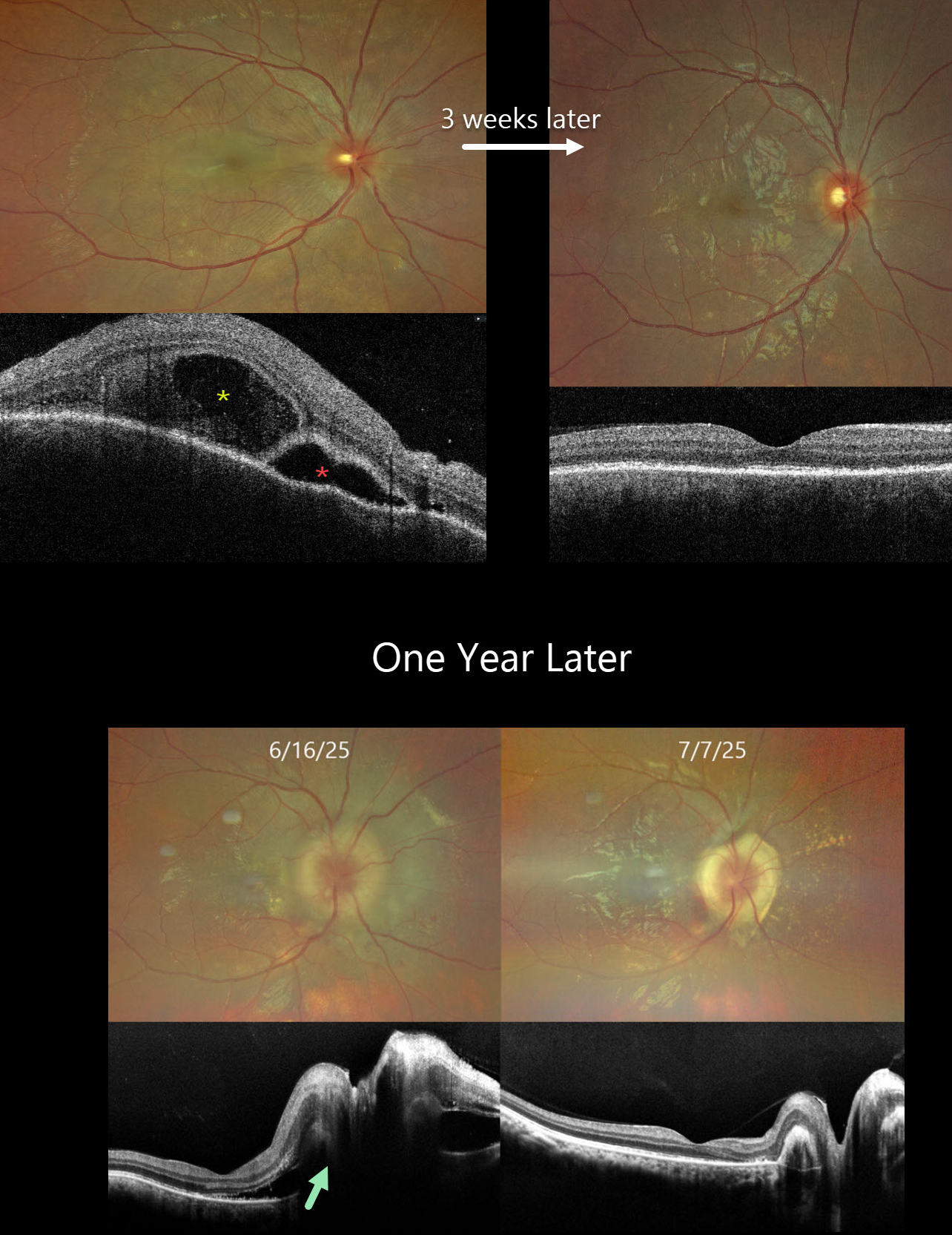
This previously healthy 24YO male presented with acute vision loss in his right eye. Vision was 20/200 OD and 20/20 in his normal OS. There were no neurologic or systemic symptoms.
Color photography shows an exudative retinal detachment involving the posterior pole. OCT shows a bacillary layer detachment, subretinal fluid with thickened retina and choroid. He was diagnosed with unilateral Harada disease and started on oral prednisone. Three weeks later, vision improved to 20/25 OD with marked clinical improvement. The oral prednisolone was tapered weekly over the next 6 weeks and stopped.
He returned one year later with recurrent vision loss. Vision was 20/200 OD. Exudative fluid with distal lipid flecks and a few small retinal hemorrhages surrounded the right nerve. OCT confirmed marked disk and peripapillary thickening with subretinal fluid. Oral prednisone was restarted, and oral azathioprine therapy was added. Three weeks later, the vision improved to 20/30. The fluid completely resolved with a ring of subretinal fibrosis surrounding the disc. The oral prednisone was tapered weekly over the next 6 weeks, and the oral azathioprine is planned to continue for the next year with regular monitoring.
Learning Points:
Harada disease is characterized by relatively acute bilateral panuveitis, optic nerve swelling, thickened choroid, chorioretinal folds, multifocal serous exudative retinal detachments, and bacillary layer detachments. Patients with just ocular findings have Harada disease, whereas those with additional systemic findings (including vitiligo, poliosis, headache, vertigo, and hearing loss) have Vogt-Koyanagi-Harada (VKH) disease. Although classically bilateral, early stages may present unilaterally before involving the fellow eye.
The disease generally progresses through four clinical stages: Prodromal, resembling a viral illness with systemic symptoms (fever, headache, meningismus); Uveitic, characterized by blurred vision, photophobia, optic disc edema, posterior choroidal thickening, and multifocal serous retinal detachments; Chronic/Convalescent, where depigmentation of the choroid leads to a “sunset-glow fundus” along with cutaneous signs like vitiligo, poliosis, and alopecia; and Recurrent, marked by repeated bouts of anterior uveitis and complications such as cataract, glaucoma, or macular neovascularization.
Management centers on high-dose systemic corticosteroids, often followed by steroid-sparing immunosuppressive agents to control inflammation and prevent recurrences. Early, aggressive therapy improves visual outcomes.



