

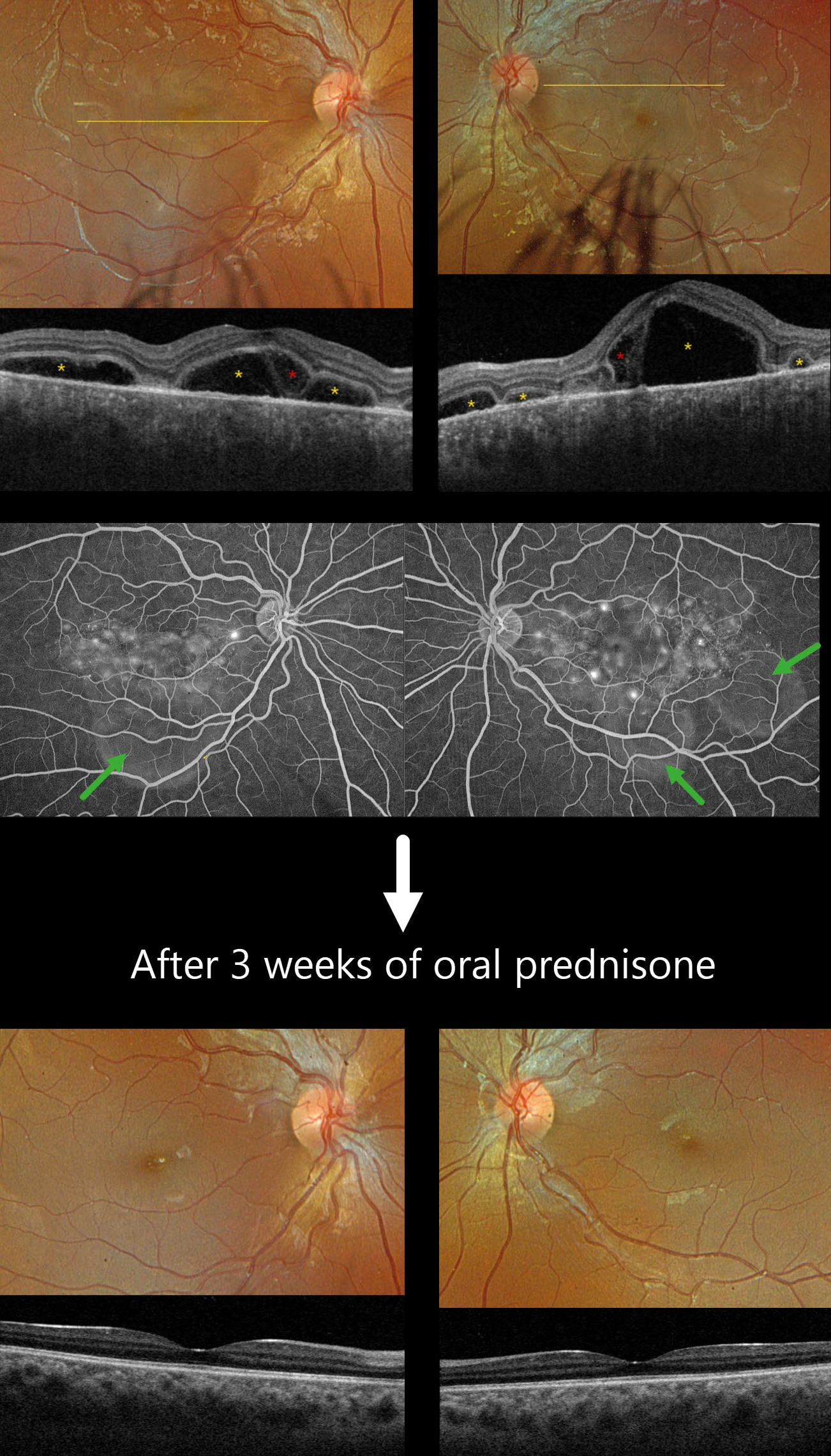
This previously healthy 13YO girl presented with several days of bilateral vision loss. Vision was 20/100 OD and 20/400 OS.
Optos color RGB imaging shows bilateral multifocal serous exudative retinal detachments. Triton swept-source OCT shows a complex pattern of multifocal pockets of subretinal fluid and bacillary layer detachments (BALAD). The choroid is markedly thickened. Fluorescein angiography shows multifocal pinpoint areas of subretinal leakage with pooling in the subretinal space and BALADs.
Learning Points:
Harada disease is characterized by relatively acute bilateral panuveitis, optic nerve swelling, thickened choroid, chorioretinal folds, multifocal serous exudative retinal detachments, and bacillary layer detachments. Patients with just ocular findings have Harada disease, whereas those with additional systemic findings (including vitiligo, poliosis, headache, vertigo, and hearing loss) have Vogt-Koyanagi-Harada disease.
Our patient was started on oral prednisone 40mg daily, and one week later, vision improved to 20/60 OU with resolution of all exudation (not shown). Three weeks later, the fundus appeared normal, and the choroid was approaching normal thickness. A slow steroid taper was started.



