

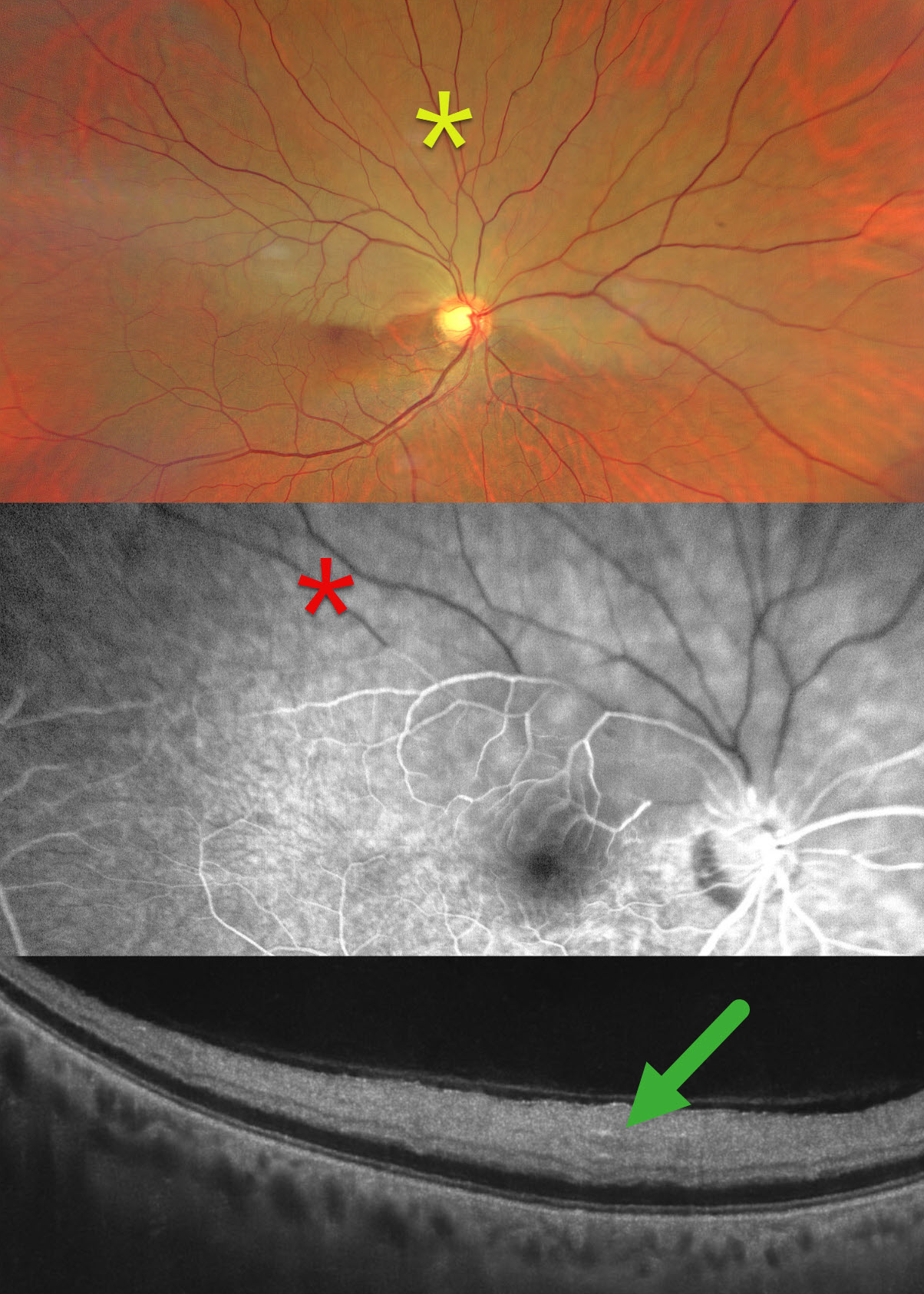
This healthy 65YO male presented with 2 days of sudden painless decreased vision in his right eye. Vision was 20/80 OD and 20/20 in his healthy OS.
Color photography shows an acute superior hemi-central retinal artery occlusion (CRAO) with opaque white retina. On fluorescein angiography, there is a marked absence of perfusion in the superior hemiretina, which persists 4 minutes into the study. On OCT, the retina is thickened and hyperreflective from the outer plexiform layer anterior to the internal limiting membrane.
Our patient was immediately referred to cardiology for a stroke protocol evaluation.
Learning Points:
Once the central retinal artery passes anteriorly through the lamina cribrosa, it branches into superior and inferior branches, which then further subdivide to supply the inner two-thirds of the neurosensory retina. Hemi-CRAO is a rare variant of CRAO caused by the blockage of one of these superior or inferior branches. As with more typical CRAO, emboli or giant cell arteritis are the most common etiologies.



