

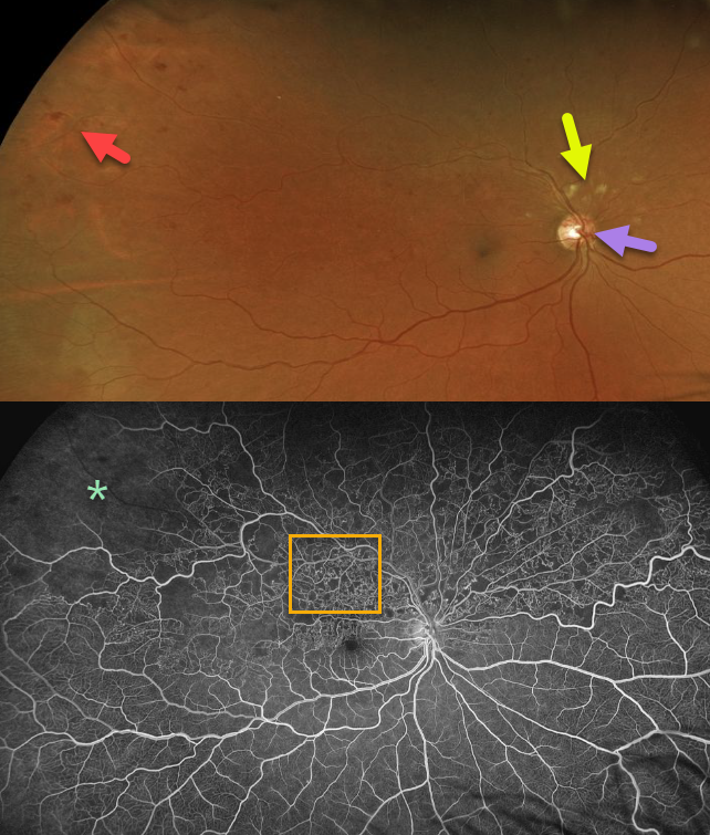
This patient has a superior hemi-reinal vein occlusion with superotemporal intraretinal hemorrhages and cotton wool spots superior to the optic nerve.
The fluorescein angiogram (FA) shows extensive retinal vascular telangiectasia and peripheral ischemia, indicating that this patient is at risk for developing retinal
neovascularization.
Learning Points:
Collateral vessels of the optic nerve can develop with retinal vein occlusions. Prominent optic nerve collateral vessels are seen in the fundus photo. These don’t leak angiographically proving that they’re not neovascular.



