

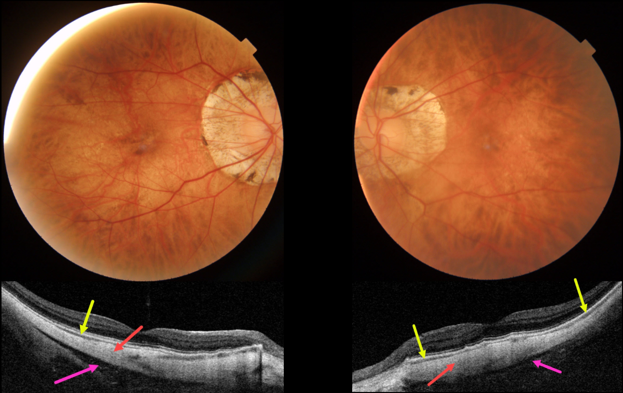
This 63YO female with severe high myopia underwent bilateral cataract surgery many years ago, along with successful vitrectomy for retinal detachment in her right eye. Vision was 20/50 OD and 20/30 OS.
Color imaging shows bilateral myopic peripapillary atrophy and macular pigmentary loss. Swept-source OCT shows a dramatically thinned or absent choroid and full-thickness, thinned sclera measuring about 300 microns bilaterally (normal posterior scleral thickness should be about 1mm). A hyporeflective region behind the sclera likely represents Tenon’s capsule, with the orbit visible more posteriorly.
Learning Points:
The era of ocular OCT began in 1991 with the initial report from Dr. Fujimoto’s lab at MIT (Huang et al, Science 1991;254:1178-1181). The technology was acquired by Zeiss in 1994, and after several failed commercial iterations, the Zeiss Stratus time-domain OCT in 2002 made it a must-have instrument for eye care.
Spectral domain units became available in 2008, followed by swept source units in 2015. Each subsequent generation can capture more information at greater retinal/choroidal depth. The swept source technology, in particular, is best able to visualize choroidal detail.
Our patient’s axial myopia with thinned choroid and sclera, along with the swept-source technology, provides a unique ability to visualize Tenon’s capsule and the anterior orbit.
For a wonderful historical review of the development, commercialization, and impact of OCT, see Fujimoto and Swanson, Invest Ophthalmol Vis Sci 2016;57;OCT1-OCT13.



