

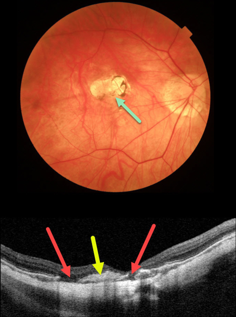
This 48YO female has a history of a previously treated myopic macular neovascularization (MNV) in her right eye, which has remained stable with observation for years. Triton color imaging shows an inactive atrophic foveal scar.
Swept source OCT shows an inactive hyperreflective central MNV with adjacent RPE defects. The choroid is severely thinned, consistent with her high myopia.
Learning Points:
An RPE aperture is a rare, recently described OCT finding most commonly seen in non-exudative AMD. Different from an RPE tear, these round RPE discontinuities typically develop from an avascular RPE detachment that remodels over time, resulting in atrophy of the RPE and outer retina.
This remodeling may result from regression of drusenoid material or damage to the RPE due to separation from the choriocapillaris (Querques G et al. Retina. 2016;36:S65-S72).
Some hypothesize that these lesions may represent small RPE micro-tears from underlying type 1 MNV causing increased hydrostatic pressure, leading to damage/atrophy of the RPE.
RPE apertures can also be found in patients with acquired vitelliform lesions (Falavarjani K et al, Canadian J Ophthalmol 2022;57:e91-e94).
We are not aware of a previously described case caused by myopic MNV.



