

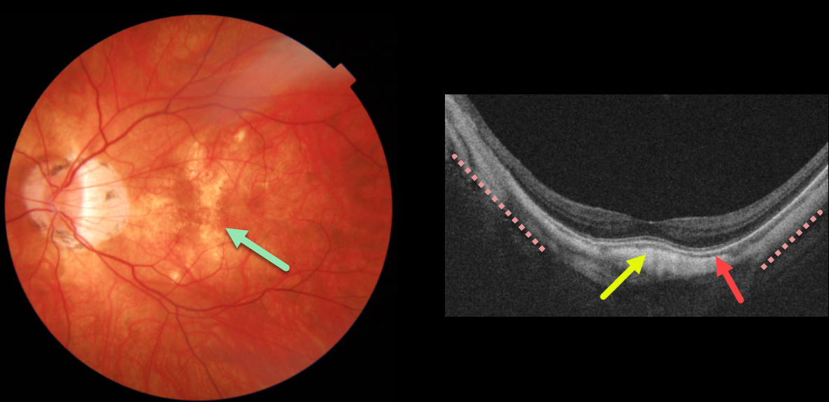
This high myope has extensive dry myopic atrophic pigmentary changes noted on Triton color imaging.
Triton swept-source OCT shows typical myopic concave posterior bowing of the eye wall, except more centrally, where there is a convex elevation beneath the fovea. The choroid is also markedly thinned throughout the posterior pole.
Learning Points:
Originally described in 2008 by Gaucher et al (AJO 2008;145:909-914), a dome-shaped macula is a macular convexity found in eyes with high myopia, often associated with a posterior staphyloma. It can be associated with avascular subretinal fluid and macular neovascularization.
See Kumar et al for an excellent recent review of this entity (Surv Ophthalmology 2021;66:560-571).



