

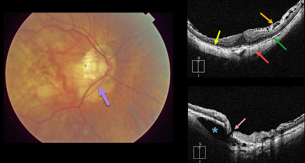
Peripapillary intrachoroidal cavitation is a relatively rare abnormality most commonly found in high myopia. Clinically, these subretinal lesions have an orange coloration and are usually located inferior to the optic nerve.
OCT scanning of the central macula shows a variety of typical myopic findings, including posterior bowing of the sclera, variable attenuation of the ellipsoid zone, markedly thinned choroid, and a variable epiretinal membrane with inner retinal schisis most prominent superiorly.
OCT scanning centered over the optic nerve reveals the intrachoroidal cavitation. There also seems to be a tiny full-thickness retinal defect that communicates directly with the cavitation.
Learning Points:
To learn more about intrachoroidal cavitations and high myopia, see Spaide et al Retina 2012;32;1037-1044.
See Ikuno Retina 2017;37;2347-2351 for a nice overview of macular findings in high myopia.



