

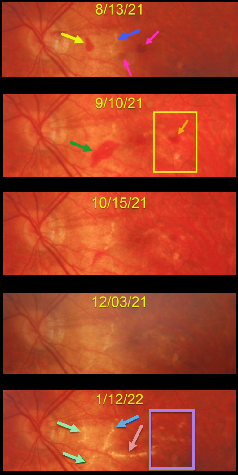
This 23YO high myopic female presented on 8/13/21 with some symptomatic central and paracentral scotomas. Vision was 20/80.
There were a few round subretinal hemorrhages near the central macula, along with an asymptomatic nasal macular hemorrhage. A few small lacquer cracks were noted.
She was then monitored monthly with an unusual rapid succession of new and resolving hemorrhages with subsequent lacquer crack development.
On 9/10/21, the prior hemorrhages were resolving, but a new area of subretinal blood was noted along the inferior edge of the previously noted lacquer crack. There was also a new hemorrhage in the distal macula. These hemorrhages all resolved by the 12/3/21 exam.
When last seen on 1/12/22, in the areas of prior subretinal blood, there are new lacquer cracks, enlargement of the originally noted lacquer crack, and a large new horizontal lacquer crack inferior to the fovea. In the region of the distal temporal macular hemorrhage noted on 9/10/21, there are numerous new lacquer cracks. Vision was 20/60, and we continue to monitor her for macular neovascularization (MNV).
Learning Points:
Lacquer cracks are a common finding in patients with high myopia. They consist of breaks in Bruch’s membrane due to the outpouching/stretching of the pathologically myopic eye and increase the risk for MNV.
In some cases, similar to this patient, they can have small avascular subretinal hemorrhages, not associated with neovascularization, and are monitored. These are best confirmed with fluorescein angiography. In cases with an active membrane, they are treated with anti-VEGF injections.



