

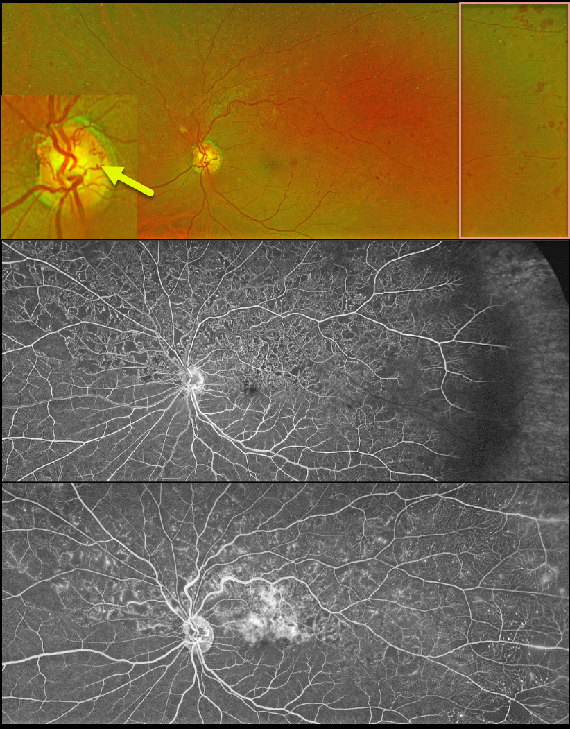
This 63YO male was referred for relatively asymptomatic retinal findings in his left eye. Vision was 20/60.
Optos color RG imaging shows a superior hemiretinal vein occlusion (HRVO) with mild hemorrhages, mostly in the temporal periphery. The occlusion is well-perfused without retinal neovascularization on fluorescein angiography. Prominent collateral vessels are noted on the optic disc superiorly. Observation was recommended.
Learning Points:
Branch retinal vein occlusions are virtually always caused by a retinal artery compressing a retinal vein within its common adventitial sheath. Some people are born with an anomalous two-trunked central retinal vein that independently drains the superior and inferior retinal hemispheres.
HRVO, similar to central retinal vein occlusion, is caused by compression of the retinal vein posterior to the lamina cribrosa (see Hayreh, Arch Ophthalmol 1980;98:1600-1609).
Collateral vessels are dilated, pre-existing vessels that bypass an occlusion. These are seen in the posterior pole most commonly following retinal venous occlusions, but can also occur on the optic disc with optic nerve tumors. The endothelial tight junctions are intact, so they don’t leak on angiography. This helps differentiate them from neovascularization, which does leak.



