

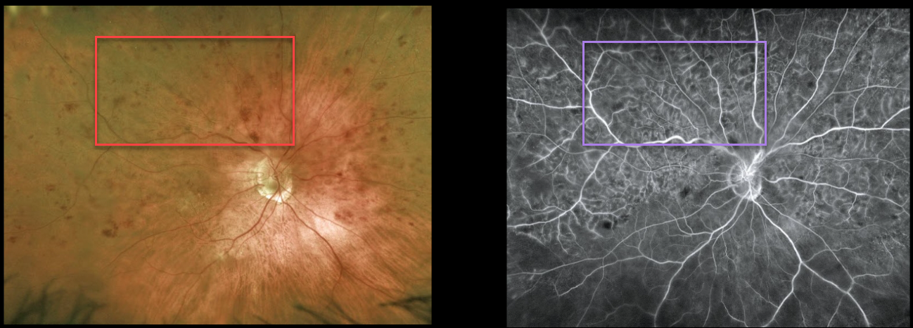
This 88YO female presented with counting fingers vision in her right eye from a symptomatic superior hemiretinal vein occlusion (HRVO).
Optos imaging shows moderate superior hemispheric inner retinal hemorrhages, and fluorescein angiography shows a well-perfused occlusion. OCT scanning showed moderate central macular edema (not shown). Anti-VEGF therapy was started.
Learning Points:
Branch retinal vein occlusions are virtually always caused by a retinal artery compressing a retinal vein within its common adventitial sheath. Some people are born with an anomalous two-trunked central retinal vein that independently drains the superior and inferior retinal hemispheres.
HRVO, similar to central retinal vein occlusion, is caused by compression of the retinal vein posterior to the lamina cribrosa (see Hayreh, Arch Ophthalmol 1980;98:1600-1609).



