

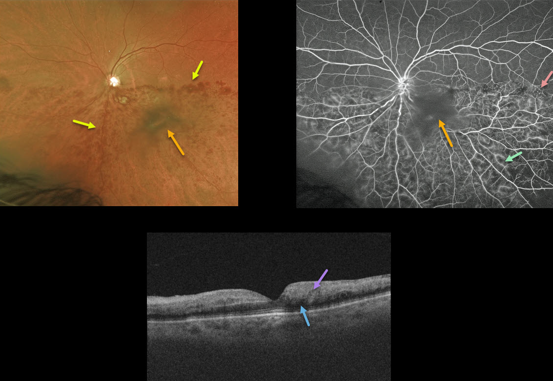
This 78YO female presented with diffuse intraretinal hemorrhages in the inferior half of her posterior pole from an inferior hemiretinal vein occlusion. She had a long history of uncontrolled hypertension, although her blood pressure had not been acutely elevated.
Fluorescein angiography shows good perfusion, some areas of vascular staining/leakage, and some telangiectasia at the border of the occlusion temporally. The large central blob in both the Optos image and the angiogram is clinically noted as a dense vitreous condensation, not blood.
OCT shows no macular edema with mild outer retinal shadowing due to the hyperreflective inner retina. Since there is no edema, we are currently observing the patient and are hoping the vision will improve as the retinal blood absorbs.
Learning Points:
Over time, patients can develop retinal atrophy from ischemic BRVO. In addition, collateral vessels are a common finding seen in venous occlusions.



