

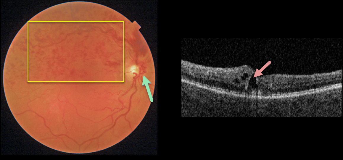
This 62YO female presented with 20/30 vision from a symptomatic superior hemiretinal vein occlusion (HRVO). Fundus imaging shows moderate retinal hemorrhages in the superior retinal hemisphere and a prominent collateral vessel along the nasal aspect of the optic nerve. There is superior macular edema on OCT.
Learning Points:
Branch retinal vein occlusions (BRVO) are virtually always caused by a retinal artery compressing a retinal vein within its common adventitial sheath. Some people are born with an anomalous two-trunked central retinal vein that independently drains the superior and inferior retinal hemispheres.
HRVO, similar to central retinal vein occlusion, is caused by compression of the retinal vein posterior to the lamina cribrosa (see Hayreh, Arch Ophthalmol 1980;98:1600-1609).
Collateral vessels are dilated, pre-existing vessels that bypass an occlusion. These are most commonly seen in the posterior pole following retinal venous occlusions, but can also occur on the optic disc with optic nerve meningiomas.
The endothelial tight junctions are intact, so there’s no leakage on angiography. This helps differentiate them from neovascularization, which does leak.



