

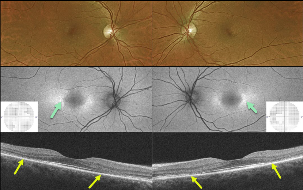
This 43YO female presented without visual complaint and 20/25 vision bilaterally. She gave a 27-year history of Plaquenil use for lupus.
Optos color imaging shows subtle pigment loss surrounding each fovea. Fundus autofluorescence (FAF) shows more obvious changes with perifoveal hyper-FAF nasally and temporally. 10-2 visual fields were normal.
Swept-source OCT B-scan imaging shows classic pericentral outer retinal atrophy.
Learning Points:
Current screening guidelines should make Plaquenil toxicity a thing of the past. Last revised in 2016, the American Academy of Ophthalmology (AAO) recommends that a baseline exam be performed before starting the medication, with annual screenings beginning at least 5 years after initiating treatment, unless major risk factors are present.
However, in practice, patients are usually screened yearly once they are placed on this medication.
Optical coherence tomography (OCT) is performed annually to assess outer retinal findings, including ellipsoid zone loss.
Our case emphasizes the importance of using multiple testing modalities to detect toxicity at the earliest possible stage.



