

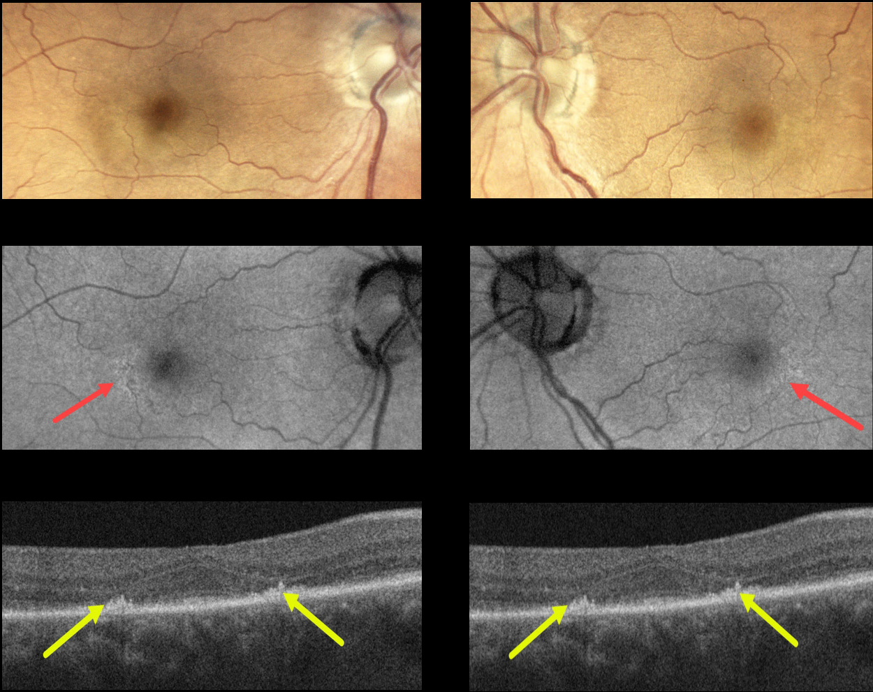
This 73YO female presented with bilateral hydroxychloroquine (Plaquenil) macular toxicity despite being treated for only 5 years. Vision was 20/40 OD and 20/30 OS.
Color photography shows very subtle pigmentary changes encircling each fovea. Fundus autofluorescence (FAF) shows subtle changes as well. The findings are most evident with OCT, which shows symmetrical paracentral outer retinal atrophy.
Learning Points:
Current screening guidelines should make Plaquenil toxicity a thing of the past. Last revised in 2016, the American Academy of Ophthalmology (AAO) recommends that a baseline exam be performed before starting the medication with annual screenings beginning at least 5 years after initiating treatment, unless major risk factors are present.
However, in practicality, patients are usually screened yearly once they are placed on this medication. Optical coherence tomography (OCT) is performed annually, looking for outer retinal findings, including ellipsoid zone loss.
Humphrey 10-2 visual field testing (24-2 for Asians since their macular involvement is usually more peripheral) is needed annually as well, since about 10% of patients will have field loss despite normal examinations and OCT testing.
Plaquenil dosing should also be based on real, not ideal, weight to better predict the optimal dose. See Marmor et al Ophthalmology 2016;123;1386-1394 for the full screening guidelines.
Our patient’s case is somewhat unique in that macular toxicity developed after only 4 years of therapy. This reinforces that screening and treatment guidelines must always be individualized.



