

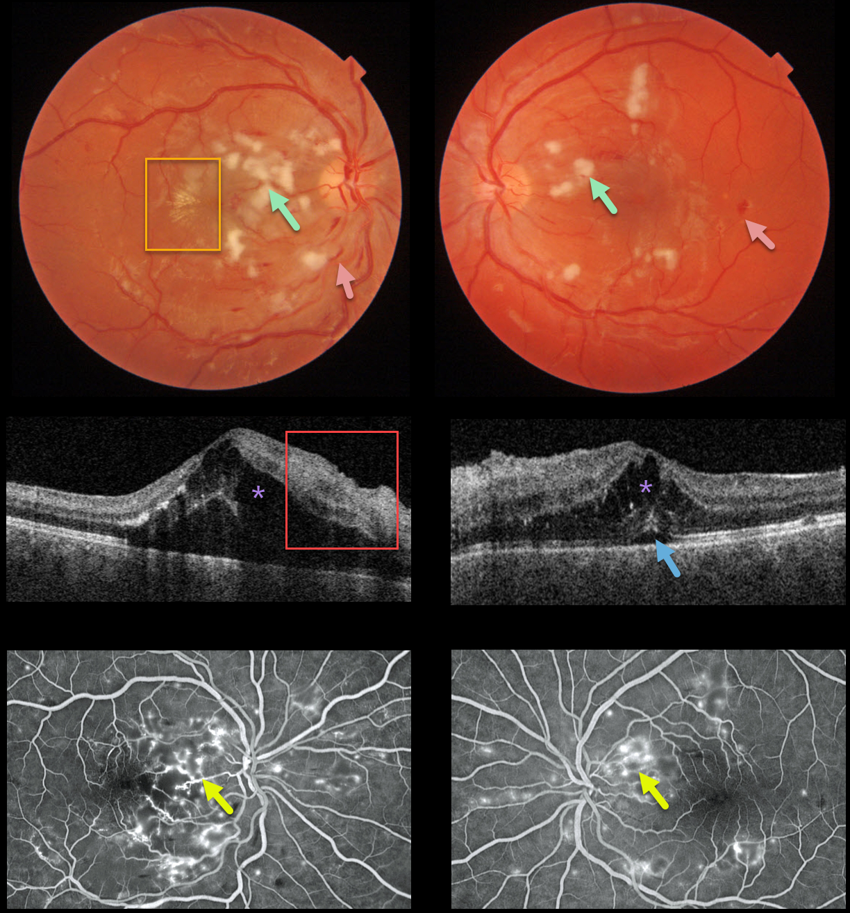
This 22YO male presented with subacute vision loss of 20/200 OD and 20/50 OS. Triton color imaging shows diffuse bilateral cotton wool spots with intraretinal hemorrhages, along with an early macular star temporal to the right fovea.
Swept-source OCT shows bilateral intraretinal thickening (mostly in the outer nuclear layer) and hyperreflective inner retinal layers, along with subfoveal fluid.
Optos fluorescein angiography shows bilateral telangiectasia and staining, mostly around the nerve fiber layer infarcts.
Blood pressure was 169/135. He was immediately sent to the nearest emergency room.
Learning Points:
Malignant hypertension, defined as blood pressure above 180 systolic and/or 120 diastolic, is a life-threatening medical emergency. Eye doctors are in a unique position to often diagnose this condition.
Patients will often present with bilateral optic nerve swelling, flame-shaped retinal hemorrhages, and, with more chronic disease, lipid precipitates in the nasal macular outer plexiform layer.
Since the macular fluid originates from the optic nerve, macular thickening is always worse nasally and is an important clinical clue for this diagnosis.



