

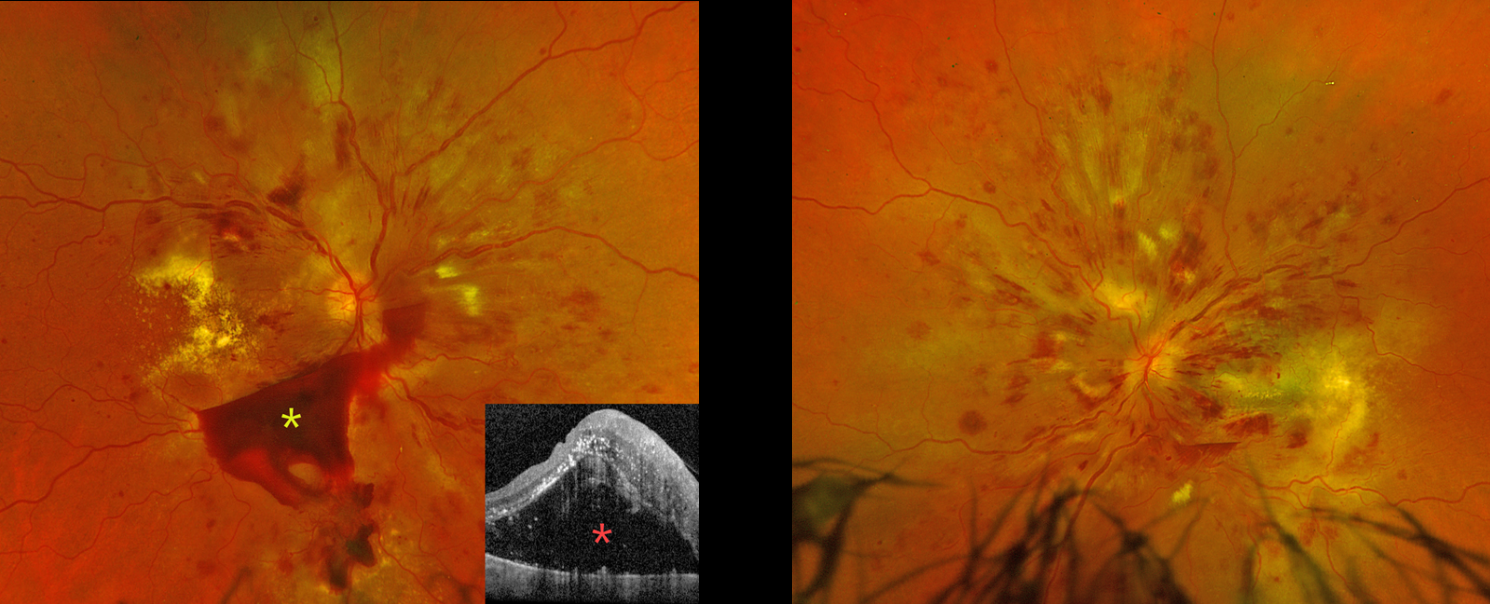
This 42YO undomiciled female presented with several months of bilateral vision loss. She has a complex medical history of hypertension, type 2 diabetes (last A1c 9.9), end-stage renal disease on dialysis, chronic obstructive pulmonary disorder, and bipolar disorder. The day prior to presenting to our clinic, she was seen at an outside emergency department for intractable vomiting in the setting of 3 consecutively missed dialysis sessions. Her blood pressure in the ED was 199/105 after self-stopping blood pressure medications because of vomiting. She had left the ED against medical advice. Vision was counting fingers OD and hand motion OS.
Optos color RG imaging OD shows diffuse intraretinal hemorrhages, multiple subhyaloid hemorrhages, cotton-wool spots (CWS), and extensive intraretinal and subretinal exudation. OCT shows marked exudative subretinal fluid with intraretinal hyperreflectivity and thickened inner retinal layers. Similar findings are noted OS.
Learning Points:
Malignant hypertension, defined as blood pressure above 180 systolic and/or 120 diastolic, is a life-threatening medical emergency. Eye doctors are often uniquely positioned to diagnose this condition (Shukla et al, Indian J Ophthalmol 2025;73:1552-1555). Patients will often present with bilateral optic nerve swelling, flame-shaped retinal hemorrhages, CWS, and, with more chronic disease, lipid precipitates in the nasal macular outer plexiform layer.
Our patient’s funduscopic findings were more likely hypertensive than from proliferative diabetic retinopathy. She was instructed to return to the emergency room for blood pressure control, and a poor visual prognosis was discussed.



