

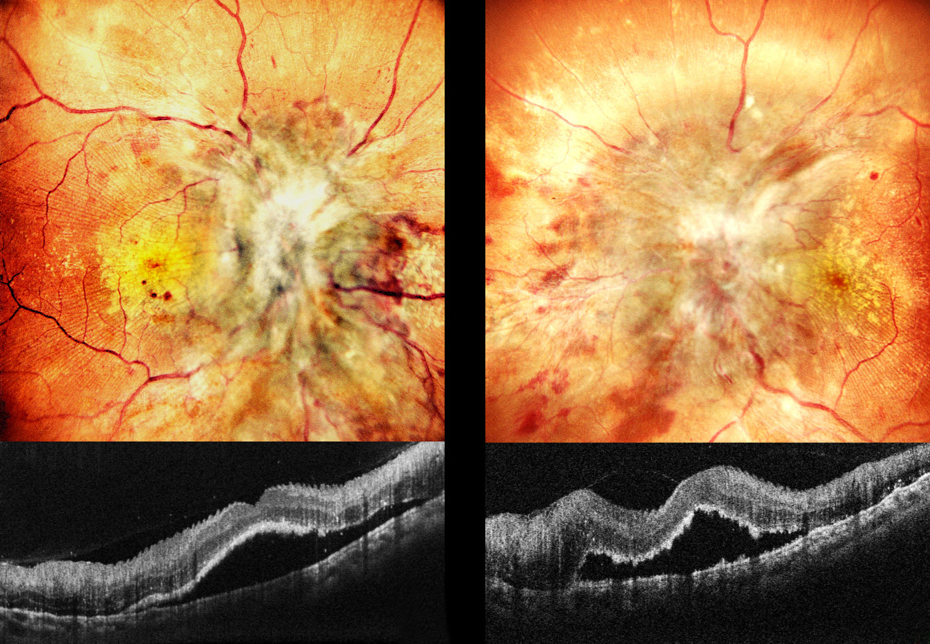
This 20YO male has a known history of chronic kidney disease. Despite hemodialysis, he’s had multiple episodes of malignant hypertension over the prior 2 months. He presented with 1 month of severe vision loss of counting fingers at 2 meters OU.
Pseudocolor SLO imaging shows profound bilateral disc edema with more peripheral nerve fiber layer infarcts (cotton-wool spots, CWS), retinal hemorrhages, and macular lipid. Fine inner retinal radiating striae are noted temporally. OCT scanning shows subfoveal fluid with hyperreflective ragged outer segments. Some small dots of outer retinal hyperreflectivity likely represent lipid.
Learning Points:
Malignant hypertension, defined as blood pressure above 180 systolic and/or 120 diastolic, is a life-threatening medical emergency. Eye doctors are often uniquely positioned to diagnose this condition (Shukla et al., Indian J Ophthalmol 2025;73:1552-1555). Patients will often present with bilateral optic nerve swelling, flame-shaped retinal hemorrhages, CWS, and, with more chronic disease, lipid precipitates in the nasal macular outer plexiform layer.
Posterior segment findings include exudative retinopathy, choroidopathy, and neuropathy. Retinopathy, due to breakdown of the inner retinal blood-retinal barrier, results in retinal hemorrhages and CWS. Choroidopathy, caused by choroidal fibrinoid necrosis, acutely creates exudative retinal detachment and, upon resolution, can leave behind generalized coarse, focal (Elschnig spots) or linear (Siegrist streaks) pigmentary changes. Papilledema is defined as bilateral disc swelling due to raised intracranial pressure. The nerve findings in malignant hypertension are likely due more to localized ischemic changes than to elevated intracranial pressure (Kishi et al, Arch Ophthalmology 1985:1198-1206).



