

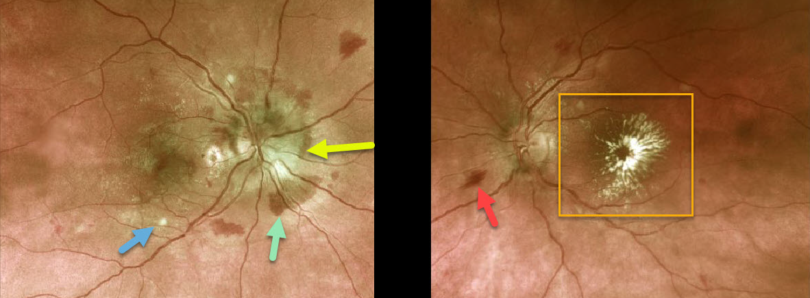
This previously healthy 36 YO female presented with recent vision loss of 20/200 bilaterally.
The right optic nerve is moderately swollen with surrounding mostly deep retinal hemorrhages, faint scattered macular lipid, and a few nerve fiber layer infarcts (cotton wool spots). The left nerve is flat with a few surrounding inner flame-shaped retinal hemorrhages and a prominent foveal lipid star.
Blood pressure was 239/159, and she was sent immediately to the emergency room for malignant hypertension.
Learning Points:
Her presentation was a bit unusual, in that systemic problems usually cause symmetric ocular findings. Her blood pressure was likely elevated for weeks to months since the macular lipid is a late finding.
We are not sure why the left nerve is not swollen, but it certainly was in the past. The lipid star is a telltale sign of prior optic nerve swelling, in which fluid and lipoproteins leaked into the surrounding tissues. When the fluid is absorbed, the lipid precipitates out in Henle’s layer. This is also seen in other causes of optic nerve swelling or inflammation, including neuroretinitis.
The deep retinal hemorrhages in her right eye are also unusual, since these are usually found in the nerve fiber layer with hypertension.



