

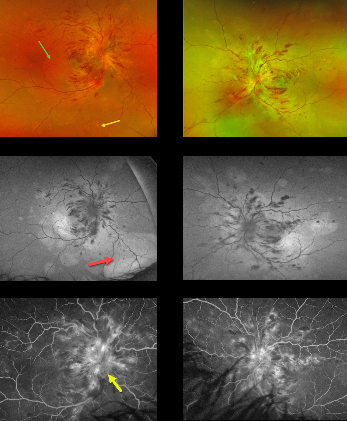
This 21YO female was admitted to the hospital 2 weeks earlier for malignant hypertension. She underwent prior failed renal transplantation due to tubulocystic renal disease and was currently on dialysis. Vision was counting fingers bilaterally.
Optos color RG imaging shows marked bilateral papilledema with mostly inner retinal hemorrhages radiating into the midperipheries. Multifocal blisters of exudative serous retinal detachments are scattered throughout each posterior pole, including the macula and midperiphery. These exudative detachments are best imaged on fundus autofluorescence (FAF), where they are hyper-FAF. Fluorescein angiography shows marked optic nerve leakage and scattered more peripheral subretinal leakage.
Learning Points:
Malignant hypertension, defined as blood pressure above 180 systolic and/or 120 diastolic, is a life-threatening medical emergency. Eye doctors are in a unique position to often diagnose this condition. Patients will often present with bilateral optic nerve swelling, flame-shaped retinal hemorrhages, and, with more chronic disease, lipid precipitates in the nasal macular outer plexiform layer.
Acute hypertensive choroidopathy outside the setting of pre-eclampsia, in our experience, is quite rare. Independent of the disc and neurosensory retinal findings, patients present with localized multifocal serous retinal detachments (de Venecia and Jampol, Arch Ophthalmol 1984;102:68-73). With blood pressure control, these resolve often with minimal funduscopic changes. Focal (Elschnig spots) or linear (Siegrist streaks) choroidal pigmentary changes may result from more severe disease. The acute findings are thought to result from choroidal arterial constriction with secondary choriocapillaris necrosis (Hayreh et al, Ophthalmology 1986;93:1383-1400). This causes ischemic injury to the overlying RPE with a breakdown of the outer blood-retinal barrier and exudative subretinal fluid.



